


1,432 results found
Case
High division of the brachial artery

Published
15 Apr 2024
75% complete
DSA (angiography)
Case
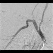
Normal brachial angiogram

Published
15 Apr 2024
29% complete
DSA (angiography)
Case
MR internal auditory canals coronal - labeling questions

Published
06 Apr 2024
40% complete
Annotated image
MRI
Case
MR internal auditory canals sagittal - labeling questions

Published
06 Apr 2024
40% complete
Annotated image
MRI
Case
MR internal auditory canals axial - labeling questions

Published
06 Apr 2024
40% complete
Annotated image
MRI
Case
Normal wrist extensor compartments US

Published
28 Mar 2024
25% complete
Ultrasound
Case
Normal carpal tunnel US

Published
28 Mar 2024
25% complete
Ultrasound
Case
Normal finger US

Published
28 Mar 2024
29% complete
Ultrasound
Case
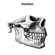
Dentition (Gray's illustrations)
Published
28 Mar 2024
35% complete
Diagram
Case
External ear (Gray's illustrations)

Published
28 Mar 2024
32% complete
Diagram
Case
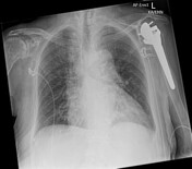
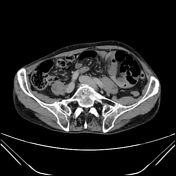
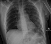
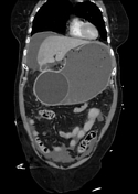
Penetrating traumatic gastric and diaphragm laceration

Published
28 Mar 2024
92% complete
CT
Annotated image
Case
Subcutaneous glucose monitor and implanon

Published
28 Mar 2024
82% complete
X-ray
Case
C6/7 chalkstick fracture in ankylosing spondylitis with large epidural hematoma

Published
28 Mar 2024
95% complete
MRI
CT
X-ray
Case
Ruptured distal biceps brachii tendon

Published
28 Mar 2024
75% complete
X-ray
Case
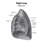
Pleural and lung margins (Gray's illustrations)

Published
28 Mar 2024
41% complete
Diagram
Case
Superior mediastinum (Gray's illustration)

Published
28 Mar 2024
32% complete
Diagram
Case
Normal subclavian angiogram
Published
28 Mar 2024
75% complete
DSA (angiography)
Case
Normal left subclavian venogram

Published
28 Mar 2024
85% complete
DSA (angiography)
Case
Normal CT humerus

Published
27 Mar 2024
33% complete
CT
Case
Normal shoulder arthrogram

Published
27 Mar 2024
72% complete
Fluoroscopy
Case
Extraocular muscles (Gray's illustration)

Published
27 Mar 2024
32% complete
Diagram
Case
Supporting structures of the eye (Gray's illustrations)

Published
27 Mar 2024
35% complete
Diagram
Case
Lacrimal apparatus (Gray's illustration)

Published
27 Mar 2024
32% complete
Diagram
Case
Tracheobronchial tree (Gray's illustration)

Published
27 Mar 2024
35% complete
Diagram
Case
Single left pulmonary vein

Published
26 Mar 2024
77% complete
CT
Case
Shepherd's crook right coronary artery (CTCA)

Published
25 Mar 2024
62% complete
CT
Case
Urinary bladder (Gray's illustration)

Published
21 Mar 2024
35% complete
Diagram
Case
Normal female pelvis - anteverted uterus (MRI)

Published
14 Mar 2024
86% complete
MRI
Case
Pseudotumor of the calcaneus

Published
12 Mar 2024
91% complete
X-ray
Case
Repeated boxers fracture of 5th metacarpal and ORIF plate

Published
07 Mar 2024
85% complete
X-ray
Case
Transabdominal pelvic US - labeling questions

Published
06 Mar 2024
36% complete
Ultrasound
Annotated image
Case
Normal CTA abdomen and pelvis (male)

Published
06 Mar 2024
48% complete
CT
Case
MR lumbar spine axial - labeling questions

Published
29 Feb 2024
40% complete
Annotated image
MRI
Case
MR lumbar spine sagittal - labeling questions

Published
29 Feb 2024
40% complete
MRI
Annotated image
Case
Pelvic fracture and hematoma due to corona mortis injury

Published
28 Feb 2024
87% complete
DSA (angiography)
CT
Case
Activated charcoal in the stomach - CT density

Published
28 Feb 2024
85% complete
CT
Case
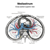
Mediastinum (Gray's illustrations)
Published
27 Feb 2024
35% complete
Diagram
Case
Pulmonary hila (Gray's illustrations)
Published
27 Feb 2024
32% complete
Diagram
Case
Ultrasound diaphragmatic sniff test

Published
26 Feb 2024
88% complete
Ultrasound
Case
Breathing artefact mimicking a sternal fracture

Published
26 Feb 2024
87% complete
CT
Annotated image
Case
Medial arm mimicking pneumothorax

Published
26 Feb 2024
85% complete
X-ray
Case
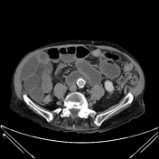
Aberrant extra renal artery and vein arising from common iliac vessels

Published
22 Feb 2024
86% complete
CT
Case
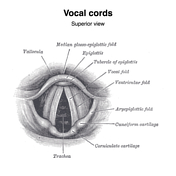
Vocal cords (Gray's illustration)
Published
20 Feb 2024
35% complete
Diagram
Case
Larynx (Gray's illustrations)

Published
20 Feb 2024
35% complete
Diagram
Case
Laryngeal muscles (Gray's illustrations)

Published
20 Feb 2024
35% complete
Diagram
Case
Laryngeal cartilages (Gray's illustrations)

Published
20 Feb 2024
35% complete
Diagram
Case
Larynx coronal CT anatomy

Published
20 Feb 2024
33% complete
Annotated image
CT
Case
Normal CTA head

Published
18 Feb 2024
59% complete
CT
Case
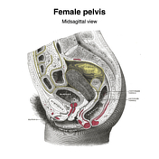
Midsagittal female pelvis (Gray's illustration)
Published
18 Feb 2024
35% complete
Diagram
Case
Orbital spaces (CT)

Published
15 Feb 2024
35% complete
Annotated image
Case
Sacroiliac erosions and subperiosteal resorption from renal osteodystrophy

Published
12 Feb 2024
92% complete
CT
Case
Ipsilateral neck of femur and pubic rami fractures

Published
08 Feb 2024
85% complete
X-ray
Case
Type 1 C2 odontoid fracture

Published
08 Feb 2024
92% complete
CT
Case
Aortic root anatomy (CTA)

Published
31 Jan 2024
35% complete
Annotated image
Case
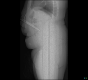
Per-oral esophageal myotomy (chest x-ray)

Published
23 Jan 2024
91% complete
X-ray
Case
Liver laceration from laparoscopic port insertion

Published
18 Jan 2024
92% complete
CT
Case
Napkin ring (photo)

Published
18 Jan 2024
35% complete
Photo
Case
Severe tracheal narrowing due to multinodular goiter following FNA

Published
03 Jan 2024
92% complete
CT
X-ray
Case
Paget disease of the rib

Published
03 Jan 2024
82% complete
X-ray
Case
Bronchopleural fistula secondary to sternoclavicular joint septic arthiritis

Published
03 Jan 2024
92% complete
X-ray
CT
Case
Failure of pubic symphysis internal fixation
Published
28 Dec 2023
89% complete
X-ray
CT
Case
Colovesical fistula due to acute sigmoid diverticulitis

Published
28 Dec 2023
95% complete
CT
Case
Cortical necrosis post-AMI and cardiac arrest
Published
28 Dec 2023
86% complete
CT
Case
Rotated dislocated glenoid component of reverse shoulder arthroplasty
Published
28 Dec 2023
91% complete
X-ray
Case
Sternomanubrial dislocation (diagrams)

Published
27 Dec 2023
44% complete
Diagram
Case
Sternomanubrial dislocation (type 1) causing buffalo pneumothorax

Published
26 Dec 2023
92% complete
X-ray
CT
Case
Traumatic elbow hemarthrosis (CT)

Published
19 Dec 2023
89% complete
CT
Case
Paronychia

Published
14 Dec 2023
88% complete
X-ray
Case
Lunate replacement

Published
12 Dec 2023
85% complete
X-ray
Case
SBO due to paraduodenal hernia
Published
06 Dec 2023
95% complete
CT
Case
Wrist arthroplasty

Published
06 Dec 2023
85% complete
X-ray
Case
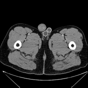
Post traumatic fat DVT and pulmonary fat embolism syndrome
Published
06 Dec 2023
95% complete
X-ray
CT
Case
Doppler effect (diagrams)

Published
30 Nov 2023
22% complete
Diagram
Case
Normal CT hand

Published
28 Nov 2023
74% complete
CT
Case
Gastric balloon causing gastric outlet obstruction and perforation
Published
20 Nov 2023
95% complete
CT
Case
Perforated Meckel's diverticulum
Published
20 Nov 2023
90% complete
Pathology
CT
Case
Penile implant and artificial urethral sphincter
Published
20 Nov 2023
95% complete
CT
Case
Shoulder resurfacing prosthesis

Published
16 Nov 2023
82% complete
X-ray
Case
Femoral pseudolesion due to penis

Published
16 Nov 2023
85% complete
X-ray
Case
Ovarian and Fallopian tube torsion
Published
05 Nov 2023
91% complete
Ultrasound
Case
Transvaginal tape
Published
05 Nov 2023
79% complete
X-ray
Case
Post-polypectomy coagulation syndrome
Published
05 Nov 2023
77% complete
CT
Case
Extensive pneumatization of the temporal bones
Published
23 Oct 2023
83% complete
CT
Case
Massive cavernous ICA aneurysm
Published
23 Oct 2023
92% complete
CT
Case
Cecal adenocarcinoma causing perforated appendicitis and ileal obstruction
Published
23 Oct 2023
86% complete
Pathology
CT
Case
Closed loop small bowel obstruction
Published
18 Oct 2023
95% complete
CT
Case
Cerebral abscess secondary to mastoiditis
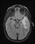
Published
18 Oct 2023
92% complete
MRI
Case
Bilateral testicular prostheses
Published
08 Oct 2023
86% complete
CT
Case
Knife penetrating abdomen indenting the IVC
Published
08 Oct 2023
85% complete
CT
Case
Recurrent laryngeal nerve palsy due to hilar lung cancer
Published
08 Oct 2023
95% complete
X-ray
CT
Case
Lingual tonsillitis
Published
08 Oct 2023
80% complete
CT
Case
Ruptured renal angiomyolipoma causing pleuritic chest pain diagnosed on CTPA

Published
08 Oct 2023
92% complete
DSA (angiography)
CT
Case
Peritoneal to abdominal height ratio
Published
28 Sep 2023
32% complete
Annotated image
Case
Aspergilloma
Published
18 Sep 2023
77% complete
X-ray
CT
ADVERTISEMENT: Supporters see fewer/no ads