


413 results found
Case
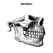
Dentition (Gray's illustrations)
Published
28 Mar 2024
35% complete
Diagram
Case
External ear (Gray's illustrations)

Published
28 Mar 2024
32% complete
Diagram
Case
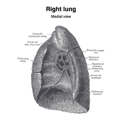
Pleural and lung margins (Gray's illustrations)

Published
28 Mar 2024
41% complete
Diagram
Case
Superior mediastinum (Gray's illustration)

Published
28 Mar 2024
32% complete
Diagram
Case
Extraocular muscles (Gray's illustration)

Published
27 Mar 2024
32% complete
Diagram
Case
Supporting structures of the eye (Gray's illustrations)

Published
27 Mar 2024
35% complete
Diagram
Case
Lacrimal apparatus (Gray's illustration)

Published
27 Mar 2024
32% complete
Diagram
Case
Tracheobronchial tree (Gray's illustration)

Published
27 Mar 2024
35% complete
Diagram
Case
Urinary bladder (Gray's illustration)

Published
21 Mar 2024
35% complete
Diagram
Case
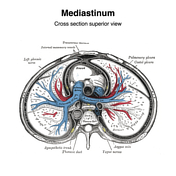
Mediastinum (Gray's illustrations)
Published
27 Feb 2024
35% complete
Diagram
Case
Pulmonary hila (Gray's illustrations)
Published
27 Feb 2024
32% complete
Diagram
Case
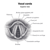
Vocal cords (Gray's illustration)
Published
20 Feb 2024
35% complete
Diagram
Case
Larynx (Gray's illustrations)

Published
20 Feb 2024
35% complete
Diagram
Case
Laryngeal muscles (Gray's illustrations)

Published
20 Feb 2024
35% complete
Diagram
Case
Laryngeal cartilages (Gray's illustrations)

Published
20 Feb 2024
35% complete
Diagram
Case
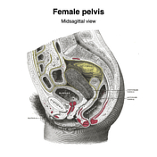
Midsagittal female pelvis (Gray's illustration)
Published
18 Feb 2024
35% complete
Diagram
Case
Sternomanubrial dislocation (diagrams)

Published
27 Dec 2023
44% complete
Diagram
Case
Doppler effect (diagrams)

Published
30 Nov 2023
22% complete
Diagram
Case
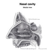
Nasal cavity (Gray's illustrations)
Published
03 May 2023
32% complete
Diagram
Case
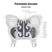
Paranasal sinuses (Gray's illustrations)
Published
03 May 2023
32% complete
Diagram
Case
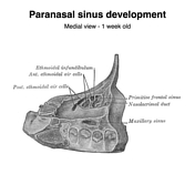
Paranasal sinus development (Gray's illustration)
Published
03 May 2023
35% complete
Diagram
Case
Nasal cartilages (Gray's illustrations)
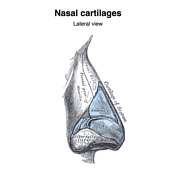
Published
03 May 2023
35% complete
Diagram
Case
Globe internal structure (Gray's illustration)
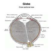
Published
03 May 2023
32% complete
Diagram
Case
Glenoid version measurement - Friedman method (diagram)
Published
21 Dec 2022
44% complete
Diagram
Case
Sympathetic nerves (Gray's illustrations)

Published
13 Oct 2022
35% complete
Diagram
Case
Autonomic ganglia of the head and neck (Gray's illustrations)

Published
13 Oct 2022
35% complete
Diagram
Case
Autonomic nervous system (Gray's illustration)

Published
12 Oct 2022
35% complete
Diagram
Case
Sacral plexus (Gray's illustrations)

Published
11 Oct 2022
35% complete
Diagram
Case
Leg and foot nerves (Gray's illustrations)

Published
11 Oct 2022
35% complete
Diagram
Case
Lower limb nerves (Gray's illustrations)

Published
10 Oct 2022
35% complete
Diagram
Case
Lumbar plexus (Gray's illustrations)

Published
06 Oct 2022
32% complete
Diagram
Case
Upper limb nerves (Gray's illustrations)

Published
02 Feb 2022
35% complete
Diagram
Case
Thoracic cutaneous nerves (Gray's illustration)

Published
02 Feb 2022
32% complete
Diagram
Case
Palmar nerves (Gray's illustrations)
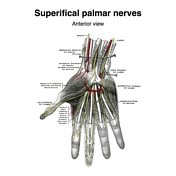
Published
27 Jan 2022
35% complete
Diagram
Case
Intercostal nerves (Gray's illustrations)

Published
27 Jan 2022
32% complete
Diagram
Case
Nerves of the face, scalp and neck (Gray's illustration)

Published
05 Jan 2022
29% complete
Diagram
Case
Cervical plexus (Gray's illustrations)

Published
05 Jan 2022
32% complete
Diagram
Case
Hypoglossal nerve (Gray's illustration)

Published
05 Jan 2022
32% complete
Diagram
Case
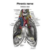
Phrenic nerve (Gray's illustration)
Published
05 Jan 2022
32% complete
Diagram
Case
Brachial plexus (Gray's illustrations)

Published
05 Jan 2022
32% complete
Diagram
Case
Suboccipital nerves (Gray's illustration)

Published
05 Jan 2022
35% complete
Diagram
Case
Posterior sacral nerves (Gray's illustration)

Published
05 Jan 2022
32% complete
Diagram
Case
Spinal nerve roots (Gray's illustrations)

Published
05 Jan 2022
32% complete
Diagram
Case
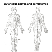
Dermatomes (Gray's illustrations)
Published
05 Jan 2022
35% complete
Diagram
Case
Cutaneous spinal nerves of the upper limb (Gray's illustrations)

Published
05 Jan 2022
29% complete
Diagram
Case
Cutaneous spinal nerves of the lower limb (Gray's illustrations)

Published
05 Jan 2022
29% complete
Diagram
Case
Origins of the extraocular muscles (Gray's illustration)

Published
27 Dec 2021
32% complete
Diagram
Case
Internal features of the lateral ventricles (Gray's illustrations)

Published
27 Dec 2021
32% complete
Diagram
Case
Innervation of the medial and lateral recti muscles (Gray's illustration)

Published
27 Dec 2021
35% complete
Diagram
Case
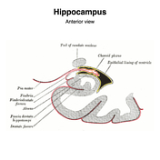
Hippocampus (Gray's illustration)
Published
27 Dec 2021
32% complete
Diagram
Case
Tela choroidea and choroid plexus of lateral ventricles (Gray's illustration)

Published
27 Dec 2021
44% complete
Diagram
Case
Internal capsule fibers (Gray's illustration)

Published
27 Dec 2021
32% complete
Diagram
Case
Fornix (Gray's illustration)

Published
27 Dec 2021
32% complete
Diagram
Case
Corpus striatum (Gray's illustration)

Published
27 Dec 2021
35% complete
Diagram
Case
Corona radiata (Gray's illustration)

Published
27 Dec 2021
32% complete
Diagram
Case
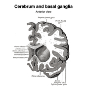
Basal ganglia (Gray's illustrations)
Published
27 Dec 2021
32% complete
Diagram
Case
Pelvic veins (Gray's illustration)
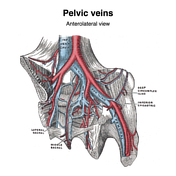
Published
20 Dec 2021
32% complete
Diagram
Case
Superficial abdominal wall veins (Gray's illustration)

Published
20 Dec 2021
29% complete
Diagram
Case
Neck veins (Gray's illustrations)

Published
20 Dec 2021
35% complete
Diagram
Case
Dural venous sinuses (Gray's illustrations)

Published
20 Dec 2021
32% complete
Diagram
Case
Veins of the scrotum (Gray's illustration)

Published
08 Dec 2021
35% complete
Diagram
Case
Female reproductive tract vessels (Gray's illustration)

Published
08 Dec 2021
35% complete
Diagram
Case
Superficial veins of the lower limb (Gray's illustration)
Published
08 Dec 2021
35% complete
Diagram
Case
Veins of the axilla (Gray's illustration)

Published
08 Dec 2021
35% complete
Diagram
Case
Tongue vessels (Gray's illustration)

Published
08 Dec 2021
35% complete
Diagram
Case
Vertebral venous plexuses (Gray's illustrations)

Published
08 Dec 2021
32% complete
Diagram
Case
Thyroid veins (Gray's illustration)

Published
08 Dec 2021
35% complete
Diagram
Case
Superficial veins of the elbow (Gray's illustration)

Published
08 Dec 2021
35% complete
Diagram
Case
Cavernous sinus (Gray's illustration)

Published
08 Dec 2021
32% complete
Diagram
Case
Superficial veins of the hand (Gray's illustration)

Published
08 Dec 2021
35% complete
Diagram
Case
Popliteal vein (Gray's illustration)
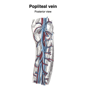
Published
08 Dec 2021
32% complete
Diagram
Case
Orbital veins (Gray's illustration)
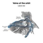
Published
08 Dec 2021
35% complete
Diagram
Case
Internal cerebral veins (Gray's illustration)

Published
07 Dec 2021
32% complete
Diagram
Case
Anal triangle (diagrams)

Published
27 Oct 2021
35% complete
Diagram
Case
Urogenital triangle (diagrams)

Published
27 Oct 2021
35% complete
Diagram
Case
Female perineal muscles (Gray's illustration)

Published
26 Oct 2021
35% complete
Diagram
Case
Male perineal muscles (Gray's illustration)

Published
26 Oct 2021
35% complete
Diagram
Case
Male perineal fascia (Gray's illustration)

Published
26 Oct 2021
29% complete
Diagram
Case
Male pelvic and perineal fascia (Gray's illustrations)

Published
26 Oct 2021
29% complete
Diagram
Case
Levator ani (Gray's illustration)

Published
26 Oct 2021
32% complete
Diagram
Case
Truncal venous development (Gray's illustrations)

Published
15 Sep 2021
35% complete
Diagram
Case
Hepatic venous development (Gray's illustration)

Published
15 Sep 2021
35% complete
Diagram
Case
Dural venous development (Gray's illustrations)

Published
15 Sep 2021
35% complete
Diagram
Case
Carotid artery development (Gray's illustration)
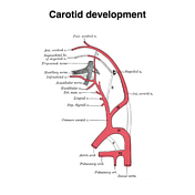
Published
15 Sep 2021
35% complete
Diagram
Case
Sinus venosus development (Gray's illustration)

Published
15 Sep 2021
29% complete
Diagram
Case
Aortic arches (Gray's illustration)

Published
14 Sep 2021
32% complete
Diagram
Case
Femoral canal (Gray's illustrations)
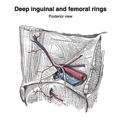
Published
29 Aug 2021
35% complete
Diagram
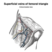
Case
Femoral triangle and sheath (Gray's illustrations)

Published
29 Aug 2021
35% complete
Diagram
Case
Internal pudendal artery (Gray's illustrations)

Published
26 Aug 2021
32% complete
Diagram
Case
Gluteal arteries (Gray's illustration)
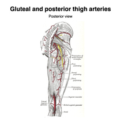
Published
26 Aug 2021
35% complete
Diagram
Case
Leg arteries (Gray's illustrations)

Published
26 Aug 2021
35% complete
Diagram
Case
Femoral artery (Gray's illustrations)

Published
26 Aug 2021
35% complete
Diagram
Case
Plantar arteries (Gray's illustrations)

Published
26 Aug 2021
32% complete
Diagram
Case
Left atrium and ventricle (Gray's illustration)

Published
26 Aug 2021
35% complete
Diagram
ADVERTISEMENT: Supporters see fewer/no ads


 Unable to process the form. Check for errors and try again.