


173 results found
Case
Radiologically inserted gastrostomy (RIG) insertion

Published
23 Apr 2024
82% complete
Fluoroscopy
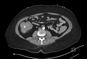
Case
Dislodged RIG with balloon deflation and pneumoperitoneum

Published
23 Apr 2024
65% complete
CT
Case
Penetrating traumatic gastric and diaphragm laceration

Published
28 Mar 2024
92% complete
CT
Annotated image
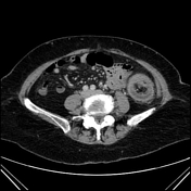
Case
Normal CTA abdomen and pelvis (male)

Published
06 Mar 2024
48% complete
CT
Case
Activated charcoal in the stomach - CT density

Published
28 Feb 2024
85% complete
CT
Case
Per-oral esophageal myotomy (chest x-ray)

Published
23 Jan 2024
91% complete
X-ray
Case
Napkin ring (photo)

Published
18 Jan 2024
35% complete
Photo
Case
Colovesical fistula due to acute sigmoid diverticulitis

Published
28 Dec 2023
95% complete
CT
Case
SBO due to paraduodenal hernia
Published
06 Dec 2023
95% complete
CT
Case
Gastric balloon causing gastric outlet obstruction and perforation
Published
20 Nov 2023
95% complete
CT
Case
Perforated Meckel's diverticulum
Published
20 Nov 2023
90% complete
Pathology
CT
Case
Post-polypectomy coagulation syndrome
Published
05 Nov 2023
77% complete
CT
Case
Cecal adenocarcinoma causing perforated appendicitis and ileal obstruction
Published
23 Oct 2023
86% complete
Pathology
CT
Case
Closed loop small bowel obstruction
Published
18 Oct 2023
95% complete
CT
Case
Knife penetrating abdomen indenting the IVC
Published
08 Oct 2023
85% complete
CT
Case
Peritoneal to abdominal height ratio
Published
28 Sep 2023
32% complete
Annotated image
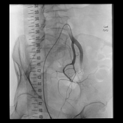
Case
Acute colonic diverticular hemorrhage requiring coil embolization
Published
11 Sep 2023
95% complete
DSA (angiography)
CT
Case
Ischemic small bowel obstruction from Morgagni hernia
Published
03 May 2023
89% complete
CT
Case
Gastric hiatus hernia
Published
03 May 2023
92% complete
X-ray
CT
Case
Small bowel obstruction due to paracaecal hernia
Published
03 May 2023
95% complete
CT
Case
Recurrent colonic cancer presenting with intussusception
Published
18 Apr 2023
80% complete
CT
Case
Post polypectomy colonic bleeding
Published
12 Jan 2023
62% complete
CT
DSA (angiography)
Case
Normal internal iliac artery angiogram
Published
16 Nov 2022
29% complete
DSA (angiography)
Case
Tooth in esophagus and stomach from intubation

Published
17 Apr 2022
90% complete
Photo
X-ray
Case
Pelvic veins (Gray's illustration)
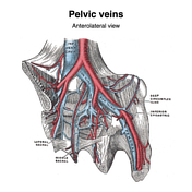
Published
20 Dec 2021
32% complete
Diagram
Case
Ischemic bowel due to internal hernia with perforation
Published
21 Nov 2021
92% complete
CT
Case
CT angiogram abdomen/pelvis sagittal - labeling questions

Published
08 Nov 2021
40% complete
Annotated image
CT
Case
Normal CTA abdomen and pelvis (female)

Published
08 Nov 2021
48% complete
CT
Case
CT angiogram abdomen/pelvis coronal - labeling questions

Published
04 Nov 2021
40% complete
Annotated image
CT
Case
CT angiogram abdomen/pelvis axial - labeling questions

Published
03 Nov 2021
40% complete
CT
Annotated image
Case
Anal triangle (diagrams)

Published
27 Oct 2021
35% complete
Diagram
Case
Urogenital triangle (diagrams)

Published
27 Oct 2021
35% complete
Diagram
Case
Female perineal muscles (Gray's illustration)

Published
26 Oct 2021
35% complete
Diagram
Case
Male perineal muscles (Gray's illustration)

Published
26 Oct 2021
35% complete
Diagram
Case
Male perineal fascia (Gray's illustration)

Published
26 Oct 2021
29% complete
Diagram
Case
Male pelvic and perineal fascia (Gray's illustrations)

Published
26 Oct 2021
29% complete
Diagram
Case
Levator ani (Gray's illustration)

Published
26 Oct 2021
32% complete
Diagram
Case
Truncal venous development (Gray's illustrations)

Published
15 Sep 2021
35% complete
Diagram
Case
Hepatic venous development (Gray's illustration)

Published
15 Sep 2021
35% complete
Diagram
Case
Femoral canal (Gray's illustrations)
Published
29 Aug 2021
35% complete
Diagram
Case
Internal pudendal artery (Gray's illustrations)

Published
26 Aug 2021
32% complete
Diagram
Case
Inguinal canal and rings (Gray's illustrations)

Published
03 Aug 2021
32% complete
Diagram
Case
Contrast filling the appendix post contrast swallow study

Published
22 Jul 2021
82% complete
X-ray
Case
Incidental large fecolith

Published
22 Feb 2021
48% complete
X-ray
CT
Case
Delayed traumatic splenic pseudoaneurysms

Published
22 Feb 2021
83% complete
CT
DSA (angiography)
Case
Lymphatics of the midgut (Gray's illustrations)

Published
25 Jan 2021
29% complete
Diagram
Case
Lymphatics of the stomach and foregut (Gray's illustrations)

Published
14 Jan 2021
22% complete
Case
Lymphatics of the colon (Gray's illustration)

Published
11 Jan 2021
32% complete
Diagram
Case
Swallowed pen

Published
03 Dec 2020
82% complete
X-ray
Case
Normal MRI pelvis (rectal cancer protocol)

Published
28 Oct 2020
83% complete
MRI
Case
Food bolus (lateral neck x-ray)

Published
05 Oct 2020
91% complete
X-ray
Case
Inferior mesenteric artery (Gray's illustration)

Published
06 Sep 2020
35% complete
Diagram
Case
Esophageal rupture post stricture dilatation

Published
01 Sep 2020
65% complete
CT
Case
Toxic megacolon

Published
01 Sep 2020
95% complete
X-ray
CT
Case
Normal common hepatic artery angiogram

Published
29 Jul 2020
29% complete
DSA (angiography)
Case
Normal inferior mesenteric artery angiogram

Published
29 Jul 2020
57% complete
DSA (angiography)
Case
Dilated esophagus due to tight gastric band

Published
28 Jul 2020
94% complete
X-ray
Case
Delayed traumatic splenic rupture

Published
14 Jul 2020
92% complete
DSA (angiography)
CT
Case
Umbilical hernia causing small bowel obstruction

Published
01 Jul 2020
89% complete
CT
Case
Small bowel obstruction due to large suprapubic incisional hernia
Published
15 Jun 2020
95% complete
X-ray
CT
Case
Splenic cleft from diaphragmatic slip
Published
10 Jun 2020
93% complete
CT
Case
Focal omental infarction

Published
10 Jun 2020
77% complete
CT
Case
Crohn disease - small bowel lesions and sacroiliac ankylosis

Published
01 Jun 2020
95% complete
CT
Case
Flipping nasogastric tube
Published
01 Jun 2020
85% complete
X-ray
Case
Normal superior mesenteric artery angiogram

Published
20 May 2020
25% complete
DSA (angiography)
Case
Acute colonic hemorrhage in ulcerative colitis

Published
14 May 2020
90% complete
CT
DSA (angiography)
Case
Mushroom (photo)
Published
23 Apr 2020
35% complete
Photo
Case
Caterpillar (photo)

Published
23 Apr 2020
29% complete
Photo
Case
Rectal foreign body - ping pong ball

Published
22 Apr 2020
85% complete
X-ray
Case
Large pancreatic pseudocyst with mass effect

Published
22 Apr 2020
89% complete
CT
Case
Cystogastrostomy stent and pigtail catheter

Published
16 Apr 2020
87% complete
X-ray
CT
Case
Traumatic duodenal perforation

Published
07 Apr 2020
92% complete
CT
Case
Esophageal perforation leak into pleural space

Published
06 Apr 2020
85% complete
Fluoroscopy
Case
Bowel ischemia with portal venous gas

Published
22 Mar 2020
92% complete
CT
Case
Morgagni hernia

Published
13 Feb 2020
88% complete
X-ray
Case
Colonic esophageal interposition

Published
29 Jan 2020
88% complete
X-ray
Case
Phantom abdominal CT

Published
14 Jan 2020
21% complete
CT
Photo
Case
Superior mediastinal mass due to esophageal duplication cyst

Published
09 Jan 2020
92% complete
CT
X-ray
Case
Massive intrathoracic stomach from chronic diaphragmatic rupture
Published
02 Jan 2020
91% complete
X-ray
Case
Pelvic question mark (Rorschach radiology)
Published
04 Dec 2019
21% complete
MRI
Case
Craniectomy bone plate in abdominal wall

Published
29 Nov 2019
83% complete
CT
Case
Small bowel obstruction on AXR

Published
27 Nov 2019
57% complete
X-ray
Case
Cystic artery anatomic variation (diagram)

Published
06 Nov 2019
38% complete
Diagram
Case
Rigler sign - bowel
Published
16 Oct 2019
94% complete
X-ray
Case
Feline esophagus

Published
01 Oct 2019
85% complete
Barium
Case
Large bowel obstruction on abdominal radiograph from colorectal cancer

Published
11 Mar 2019
88% complete
X-ray
Case
Thumbprinting

Published
05 Mar 2019
60% complete
X-ray
Case
Abdominal ratio

Published
05 Mar 2019
29% complete
Annotated image
Case
Small bowel obstruction on AXR

Published
28 Feb 2019
91% complete
X-ray
Case
Large right inguinal hernia - pelvic x-ray

Published
26 Feb 2019
91% complete
X-ray
Case
CT abdomen/pelvis sagittal - labeling questions

Published
13 Dec 2018
40% complete
CT
Annotated image
Case
CT abdomen/pelvis coronal - labeling questions

Published
04 Dec 2018
40% complete
Annotated image
CT
Case
Gastric wall thickening - linitis plastica (abdominal x-ray)

Published
26 Nov 2018
72% complete
X-ray
Case
CT abdomen/pelvis (lower) axial - labeling questions

Published
20 Nov 2018
40% complete
CT
Annotated image
ADVERTISEMENT: Supporters see fewer/no ads


 Unable to process the form. Check for errors and try again.