


96 results found
Case
Urinary bladder (Gray's illustration)

Published
21 Mar 2024
35% complete
Diagram
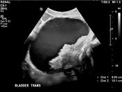
Case
Transabdominal pelvic US - labeling questions

Published
06 Mar 2024
36% complete
Ultrasound
Annotated image
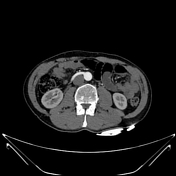
Case
Normal CTA abdomen and pelvis (male)

Published
06 Mar 2024
48% complete
CT
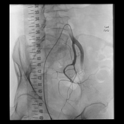
Case
Aberrant extra renal artery and vein arising from common iliac vessels

Published
22 Feb 2024
86% complete
CT
Case
Midsagittal female pelvis (Gray's illustration)
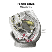
Published
18 Feb 2024
35% complete
Diagram
Case
Sacroiliac erosions and subperiosteal resorption from renal osteodystrophy

Published
12 Feb 2024
92% complete
CT
Case
Colovesical fistula due to acute sigmoid diverticulitis

Published
28 Dec 2023
95% complete
CT
Case
Penile implant and artificial urethral sphincter
Published
20 Nov 2023
95% complete
CT
Case
Bilateral testicular prostheses
Published
08 Oct 2023
86% complete
CT
Case
Ruptured renal angiomyolipoma causing pleuritic chest pain diagnosed on CTPA

Published
08 Oct 2023
92% complete
DSA (angiography)
CT
Case
Spermatic cord hematoma
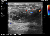
Published
11 Sep 2023
88% complete
Ultrasound
Case
VUJ calculus in ureterocoele
Published
11 Sep 2023
95% complete
X-ray
CT
Case
Bladder cancer
Published
11 Sep 2023
85% complete
Ultrasound
Case
Precaval accessory right renal artery
Published
14 Apr 2023
83% complete
CT
Case
Normal internal iliac artery angiogram
Published
16 Nov 2022
29% complete
DSA (angiography)
Case
Dual split Mirena IUDs for septate uterus

Published
11 Oct 2022
85% complete
X-ray
Case
Pelvic veins (Gray's illustration)
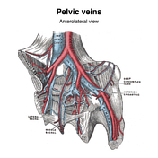
Published
20 Dec 2021
32% complete
Diagram
Case
Veins of the scrotum (Gray's illustration)

Published
08 Dec 2021
35% complete
Diagram
Case
Female reproductive tract vessels (Gray's illustration)

Published
08 Dec 2021
35% complete
Diagram
Case
CT angiogram abdomen/pelvis sagittal - labeling questions

Published
08 Nov 2021
40% complete
Annotated image
CT
Case
Normal CTA abdomen and pelvis (female)

Published
08 Nov 2021
48% complete
CT
Case
CT angiogram abdomen/pelvis coronal - labeling questions

Published
04 Nov 2021
40% complete
Annotated image
CT
Case
CT angiogram abdomen/pelvis axial - labeling questions

Published
03 Nov 2021
40% complete
CT
Annotated image
Case
Anal triangle (diagrams)

Published
27 Oct 2021
35% complete
Diagram
Case
Urogenital triangle (diagrams)

Published
27 Oct 2021
35% complete
Diagram
Case
Female perineal muscles (Gray's illustration)

Published
26 Oct 2021
35% complete
Diagram
Case
Male perineal muscles (Gray's illustration)

Published
26 Oct 2021
35% complete
Diagram
Case
Male perineal fascia (Gray's illustration)

Published
26 Oct 2021
29% complete
Diagram
Case
Male pelvic and perineal fascia (Gray's illustrations)

Published
26 Oct 2021
29% complete
Diagram
Case
Internal pudendal artery (Gray's illustrations)

Published
26 Aug 2021
32% complete
Diagram
Case
Inguinal canal and rings (Gray's illustrations)

Published
03 Aug 2021
32% complete
Diagram
Case
Bell clapper (photo)

Published
21 Jan 2021
38% complete
Photo
Case
Bell clapper deformity (diagram)

Published
21 Jan 2021
35% complete
Diagram
Case
Lymphatics of the bladder (Gray's illustration)

Published
11 Jan 2021
32% complete
Diagram
Case
Lymphatics of the prostate (Gray's illustration)

Published
11 Jan 2021
32% complete
Diagram
Case
Lymphatics of the uterus (Gray's illustration)

Published
11 Jan 2021
32% complete
Diagram
Case
Menstrual cup on trauma imaging

Published
17 Dec 2020
80% complete
X-ray
CT
Case
Anatomy of the penis (Gray's illustration)

Published
02 Sep 2020
35% complete
Diagram
Case
Venous drainage of the penis (Gray's illustration)

Published
02 Sep 2020
35% complete
Diagram
Case
Arterial supply of the penis (Gray's illustration)

Published
02 Sep 2020
35% complete
Diagram
Case
Testes and epididymis (Gray's illustration)

Published
02 Sep 2020
35% complete
Diagram
Case
Testes and spermatic cord (Gray's illustration)

Published
02 Sep 2020
35% complete
Diagram
Case
Prostate gland and seminal vesicles (Gray's illustration)

Published
02 Sep 2020
35% complete
Diagram
Case
Scrotal ring

Published
01 Sep 2020
50% complete
X-ray
Case
Renal artery stenosis on captopril renal MAG3 nuclear study
Published
30 Jul 2020
94% complete
Nuclear medicine
Case
Normal captopril renal MAG3 nuclear study

Published
29 Jul 2020
94% complete
Nuclear medicine
Case
Normal renal MAG3 nuclear study

Published
27 Jul 2020
94% complete
Nuclear medicine
Case
SBO, pancreatic and renal transplants

Published
14 Jul 2020
92% complete
CT
Case
Dual ureteric calculi

Published
24 Jun 2020
89% complete
CT
Case
Breast metastases from renal cell cancer

Published
22 Jun 2020
89% complete
Ultrasound
CT
Case
Cobra (photo)
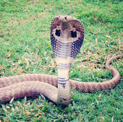
Published
23 Apr 2020
35% complete
Photo
Case
Normal scrotal ultrasound

Published
10 Mar 2020
41% complete
Ultrasound
Case
Phantom abdominal CT

Published
14 Jan 2020
21% complete
CT
Photo
Case
Paintbrush sign of medullary sponge kidney on IVP

Published
23 Dec 2019
75% complete
X-ray
Case
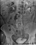
Massive bladder simulating pelvic mass on AXR
Published
23 Dec 2019
85% complete
X-ray
Case
Penile fracture grading (diagrams)

Published
22 Oct 2019
35% complete
Diagram
Case
CT abdomen/pelvis sagittal - labeling questions

Published
13 Dec 2018
40% complete
CT
Annotated image
Case
CT abdomen/pelvis coronal - labeling questions

Published
04 Dec 2018
40% complete
Annotated image
CT
Case
CT abdomen/pelvis (lower) axial - labeling questions

Published
20 Nov 2018
40% complete
CT
Annotated image
Case
CT abdomen/pelvis (upper) axial - labeling questions

Published
07 Nov 2018
40% complete
Annotated image
CT
Case
Penis anatomy (diagram)

Published
27 Sep 2018
25% complete
Diagram
Case
Humeral pathological fracture from metastasis treated with angioembolisation and IM nail

Published
14 Sep 2018
88% complete
X-ray
DSA (angiography)
Case
Normal renal tract ultrasound (female)

Published
07 Jun 2018
50% complete
Ultrasound
Case
Abdominal surface anatomy (creative commons illustration)
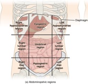
Published
20 Mar 2018
35% complete
Diagram
Case
Anatomical relations (creative commons illustration)

Published
20 Mar 2018
32% complete
Diagram
Case
Anatomical planes (creative commons illustration)
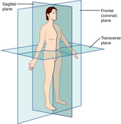
Published
20 Mar 2018
32% complete
Diagram
Case
Bladder calculus

Published
15 Mar 2018
66% complete
X-ray
Case
Renal trauma in pregnancy

Published
24 Jan 2018
94% complete
CT
Case
Mach effect (diagram)

Published
19 Oct 2017
44% complete
Diagram
Case
Bifid renal pelvis on IVP

Published
04 Oct 2017
82% complete
Fluoroscopy
Case
Abdominal aorta and retroperitoneum (Gray's illustration)

Published
17 Jul 2017
38% complete
Diagram
Case
Iliac arteries (Gray's illustration)

Published
13 Jul 2017
35% complete
Diagram
Case
Dromedary hump of the left kidney
Published
31 May 2017
66% complete
Ultrasound
Case
Retroperitoneal hemorrhage due to ruptured renal cyst in PCKD

Published
05 Jan 2017
77% complete
CT
Case
Bladder cancer in a diverticulum

Published
28 Dec 2016
74% complete
CT
Case
Radiolucent indinavir ureteric calculus

Published
07 Jan 2016
89% complete
CT
Case
Pancreatic transection with liver, renal and colonic lacerations

Published
29 Dec 2015
85% complete
CT
Case
Suprapubic catheter stuck in the urethra

Published
03 Dec 2015
89% complete
CT
Case
Normal voiding cystourethrogram

Published
01 Dec 2015
88% complete
Fluoroscopy
Case
Normal abdominal radiograph

Published
07 Nov 2015
47% complete
X-ray
Case
Extraperitoneal bladder rupture

Published
13 Oct 2015
89% complete
CT
Case
Pelviureteric junction obstruction and contralateral bifid ureter

Published
08 Sep 2015
89% complete
CT
Case
Abdominal multi-trauma - devascularised kidney and liver, spleen and pancreatic lacerations

Published
28 Aug 2015
80% complete
CT
Case
Lippe's loop (IUD) in peritoneal cavity due to previous uterine perforation

Published
28 Aug 2015
71% complete
X-ray
CT
Case
Ureteric sciatic herniation

Published
28 Aug 2015
95% complete
CT
Case
Calciphylaxis

Published
19 Aug 2015
68% complete
CT
Case
Pelvic kidney

Published
06 Aug 2015
83% complete
CT
Case
Pantaloon hernia

Published
29 Jul 2015
68% complete
CT
Case
Ureteric calculi in a horseshoe kidney

Published
28 Jul 2015
92% complete
CT
Case
Calcified occluded femoral grafts in a renal transplant patient

Published
16 Jul 2015
79% complete
X-ray
Case
Lumbar plexus (diagram)

Published
14 Jul 2015
29% complete
Diagram
Case
Transplant kidney vesicoureteric junction calculus

Published
13 Jul 2015
92% complete
CT
Case
Renal malrotation

Published
19 Jun 2015
89% complete
CT
Case
Rhabdomyolysis

Published
19 Jun 2015
92% complete
CT
ADVERTISEMENT: Supporters see fewer/no ads


 Unable to process the form. Check for errors and try again.