


1,438 results found
Case
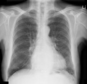
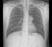
Left lower lobe pneumonia

Published
08 Nov 2019
91% complete
X-ray
Case
Acute P1 occlusion with PCA ischemia penumbra (CT perfusion)

Published
07 Nov 2019
95% complete
MRI
CT
DSA (angiography)
Case
Double density sign of left atrial enlargement

Published
06 Nov 2019
82% complete
X-ray
Case
Cystic artery anatomic variation (diagram)

Published
06 Nov 2019
38% complete
Diagram
Case
Acute ICA ischemic penumbra due to high-grade CCA stenosis (CT perfusion)

Published
05 Nov 2019
94% complete
CT
Case
Acute A3 occlusion with ACA ischemic penumbra (CT perfusion)

Published
05 Nov 2019
94% complete
CT
Case
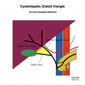
Cystohepatic (Calot) triangle (diagram)
Published
29 Oct 2019
38% complete
Diagram
Case
Acute M1 occlusion with ischemic penumbra (CT perfusion)

Published
29 Oct 2019
94% complete
DSA (angiography)
CT
Case
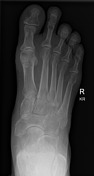
Calcified gluteal granulomata

Published
24 Oct 2019
79% complete
X-ray
Case
Penile fracture grading (diagrams)

Published
22 Oct 2019
35% complete
Diagram
Case
Polio pelvis

Published
16 Oct 2019
82% complete
X-ray
Case
Rigler sign - bowel
Published
16 Oct 2019
94% complete
X-ray
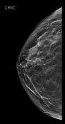
Case
Benign breast calcifications - bilateral (chest x-ray)

Published
16 Oct 2019
79% complete
X-ray
Case
Fractured left pleural catheter
Published
16 Oct 2019
90% complete
X-ray
Annotated image
Case
CABG stent

Published
08 Oct 2019
82% complete
X-ray
Case
Bilateral hilar lymphadenopathy from sarcoidosis

Published
08 Oct 2019
85% complete
X-ray
Case
Ascending aortic aneurysm
Published
08 Oct 2019
63% complete
X-ray
Case
Alveolar proteinosis

Published
01 Oct 2019
91% complete
X-ray
Case
Feline esophagus

Published
01 Oct 2019
85% complete
Barium
Case
Renal osteodystrophy

Published
22 Sep 2019
82% complete
X-ray
Case
Popliteal fossa (diagram)

Published
28 Aug 2019
29% complete
Diagram
Case
Femoral triangle (diagram)

Published
26 Aug 2019
22% complete
Diagram
Case
Cubital fossa (diagram)

Published
21 Aug 2019
29% complete
Diagram
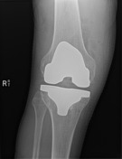
Case
Acromioclavular joint disruption treated with coracoclavicular ligament reconstruction

Published
17 Aug 2019
79% complete
X-ray
Case
Biliary duct anatomic variation (diagram)

Published
17 Aug 2019
38% complete
Diagram
Case
Pancreatic duct anatomic variation (diagram)

Published
17 Aug 2019
38% complete
Diagram
Case
Brain perfusion - time attenuation curves

Published
13 Aug 2019
29% complete
Diagram
Case
Coronary angiogram showing inflation and deflation of intra-aortic balloon pump

Published
19 Jul 2019
50% complete
DSA (angiography)
Case
Acute Stieda fracture

Published
17 Jul 2019
91% complete
X-ray
Case
Liposclerosing myxofibrous tumor

Published
17 Jul 2019
69% complete
X-ray
Case
Hand arthropathies - distribution (diagram)

Published
17 Jul 2019
16% complete
Diagram
Case
Pacemaker and brainstem stimulators (chest x-ray)

Published
28 Jun 2019
85% complete
X-ray
Case
Salter Harris IV of the proximal phalanx

Published
27 Jun 2019
82% complete
X-ray
Case
Normal calcaneum radiographs

Published
27 Jun 2019
41% complete
X-ray
Case
Left ventricular myocardial segments (diagram)

Published
02 Jun 2019
41% complete
Diagram
Case
Cantlie's line (diagram)

Published
26 May 2019
35% complete
Annotated image
Diagram
Case
Hepatectomy and sectionectomy (diagram)

Published
26 May 2019
29% complete
Diagram
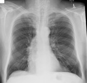
Case
Cannonball metastases - chest x-ray

Published
06 May 2019
82% complete
X-ray
Case
Subdermal magnet implant in finger

Published
06 May 2019
85% complete
X-ray
Case
Rectus sheath (diagram)

Published
10 Apr 2019
35% complete
Diagram
Case
Lateral triangular space (Gray's illustration)

Published
25 Mar 2019
29% complete
Diagram
Case
Medial triangular space (Gray's illustration)
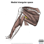
Published
25 Mar 2019
29% complete
Diagram
Case
Quadrilateral space (Gray's illustration)

Published
25 Mar 2019
32% complete
Diagram
Case
Large bowel obstruction on abdominal radiograph from colorectal cancer

Published
11 Mar 2019
88% complete
X-ray
Case
Extension teardrop fracture (x-ray)
Published
06 Mar 2019
94% complete
X-ray
Case
Thumbprinting

Published
05 Mar 2019
60% complete
X-ray
Case
Abdominal ratio

Published
05 Mar 2019
29% complete
Annotated image
Case
Unilateral facet lock

Published
04 Mar 2019
91% complete
X-ray
Case
CT temporal bone axial - labeling questions

Published
01 Mar 2019
40% complete
Annotated image
CT
Case
Small bowel obstruction on AXR

Published
28 Feb 2019
91% complete
X-ray
Case
Anatomical neck of humerus fracture

Published
27 Feb 2019
91% complete
X-ray
Case
Osteochondromata

Published
27 Feb 2019
82% complete
X-ray
Case
Iliac wing fracture
Published
27 Feb 2019
75% complete
CT
X-ray
Case
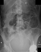
Large right inguinal hernia - pelvic x-ray

Published
26 Feb 2019
91% complete
X-ray
Case
Calcified left ventricular mural thrombus

Published
26 Feb 2019
69% complete
X-ray
Case
Flexion teardrop fracture (x-ray)
Published
26 Feb 2019
88% complete
X-ray
Case
Acute respiratory distress syndrome (ARDS)

Published
26 Feb 2019
89% complete
CT
X-ray
Case
Loculated pleural effusion on CXR

Published
26 Feb 2019
94% complete
X-ray
Case
Right mesial temporal lobe jet hematoma from ruptured PCom artery aneurysm

Published
26 Feb 2019
95% complete
CT
Case
Retrodiaphragmatic pericardial fat tag sign

Published
21 Feb 2019
91% complete
X-ray
Case
Pericardial drain on CXR

Published
21 Feb 2019
79% complete
X-ray
Case
Malpositioned intra-aortic balloon pump - too high

Published
21 Feb 2019
91% complete
X-ray
Case
Malpositioned intra-aortic balloon pump - too low

Published
21 Feb 2019
91% complete
X-ray
Case
Sternal dehiscence (on chest radiograph)

Published
21 Feb 2019
88% complete
X-ray
Case
Tripartite medial hallux sesamoid bone
Published
21 Feb 2019
85% complete
X-ray
Case
Acute pulmonary edema

Published
18 Feb 2019
72% complete
X-ray
Case
Malpositioned thermometer probe in the left main bronchus

Published
17 Feb 2019
79% complete
X-ray
Case
Pneumothorax and skin fold

Published
17 Feb 2019
91% complete
X-ray
Case
Traumatic pneumothorax - subtle (supine chest radiograph)

Published
15 Feb 2019
85% complete
X-ray
Case
Oreo cookie sign of pericardial effusion

Published
14 Feb 2019
95% complete
CT
X-ray
Annotated image
Case
Pericardial fat tag sign

Published
14 Feb 2019
92% complete
X-ray
CT
Case
Bilateral pericardial fat tag signs

Published
14 Feb 2019
66% complete
X-ray
Case
Normal MR pulmonary angiogram TWIST

Published
13 Feb 2019
45% complete
MRI
Case
Buffalo pneumothorax - post-operative

Published
07 Feb 2019
94% complete
X-ray
Case
Stove-in chest

Published
05 Feb 2019
90% complete
CT
X-ray
Case
Unfused paracondylar process

Published
30 Jan 2019
83% complete
CT
Case
Normal cervical disc replacement and anterior cervical discectomy and fusion (ACDF)

Published
11 Jan 2019
91% complete
X-ray
Case
Breast cancer workup with ultrasound hookwire localization

Published
09 Jan 2019
86% complete
Ultrasound
Mammography
Pathology
Case
Stereotactic tomosynthesis biospy of malignant breast calcifications

Published
09 Jan 2019
91% complete
Mammography
Case
Normal breast mammography (tomosynthesis) and ultrasound
Published
04 Jan 2019
83% complete
Ultrasound
Mammography
Annotated image
Case
Pneumocystis pneumonia - neonatal

Published
04 Jan 2019
94% complete
X-ray
Case
Aortic aneurysm (chest radiograph)

Published
02 Jan 2019
83% complete
X-ray
CT
Case
Fractured clavicle and ribs with pneumothorax

Published
02 Jan 2019
91% complete
X-ray
Case
Calcified loose bodies in a Baker cyst
Published
28 Dec 2018
80% complete
X-ray
CT
Case
Pubic symphysis septic arthritis and osteomyelitis

Published
27 Dec 2018
94% complete
X-ray
Case
CT abdomen/pelvis sagittal - labeling questions

Published
13 Dec 2018
40% complete
CT
Annotated image
Case
CT abdomen/pelvis coronal - labeling questions

Published
04 Dec 2018
40% complete
Annotated image
CT
Case
Anterior mediastinal mass - thymoma
Published
26 Nov 2018
92% complete
X-ray
CT
Case
Gastric wall thickening - linitis plastica (abdominal x-ray)

Published
26 Nov 2018
72% complete
X-ray
Case
Patellar tendon rupture

Published
22 Nov 2018
88% complete
X-ray
Ultrasound
Case
Acromioclavicular joint dissociation - type IV

Published
22 Nov 2018
89% complete
X-ray
CT
Case
Diaphragmatic eventration and Chilaiditi sign

Published
21 Nov 2018
85% complete
X-ray
Case
Skin fold mimicking an apical pneumothorax

Published
21 Nov 2018
88% complete
X-ray
Case
Syndesmotic injury (ankle stress view)
Published
21 Nov 2018
91% complete
X-ray
ADVERTISEMENT: Supporters see fewer/no ads


 Unable to process the form. Check for errors and try again.