


1,433 results found
Case
Haglund deformity

Published
17 Apr 2022
82% complete
X-ray
Case
Dislocated proximal and distal interphalangeal joints - hand

Published
17 Apr 2022
88% complete
X-ray
Case
Aberrant right vertebral artery with a retro-esophageal course

Published
02 Feb 2022
89% complete
CT
Case
Upper limb nerves (Gray's illustrations)

Published
02 Feb 2022
35% complete
Diagram
Case
Thoracic cutaneous nerves (Gray's illustration)

Published
02 Feb 2022
32% complete
Diagram
Case
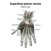
Palmar nerves (Gray's illustrations)
Published
27 Jan 2022
35% complete
Diagram
Case
Intercostal nerves (Gray's illustrations)

Published
27 Jan 2022
32% complete
Diagram
Case
Nerves of the face, scalp and neck (Gray's illustration)

Published
05 Jan 2022
29% complete
Diagram
Case
Cervical plexus (Gray's illustrations)

Published
05 Jan 2022
32% complete
Diagram
Case
Acro-osteolysis from end-stage renal failure

Published
05 Jan 2022
91% complete
X-ray
Case
Hypoglossal nerve (Gray's illustration)

Published
05 Jan 2022
32% complete
Diagram
Case
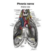
Phrenic nerve (Gray's illustration)
Published
05 Jan 2022
32% complete
Diagram
Case
Brachial plexus (Gray's illustrations)

Published
05 Jan 2022
32% complete
Diagram
Case
Suboccipital nerves (Gray's illustration)

Published
05 Jan 2022
35% complete
Diagram
Case
Posterior sacral nerves (Gray's illustration)

Published
05 Jan 2022
32% complete
Diagram
Case
Spinal nerve roots (Gray's illustrations)

Published
05 Jan 2022
32% complete
Diagram
Case
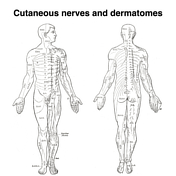
Dermatomes (Gray's illustrations)
Published
05 Jan 2022
35% complete
Diagram
Case
Cutaneous spinal nerves of the upper limb (Gray's illustrations)

Published
05 Jan 2022
29% complete
Diagram
Case
Cutaneous spinal nerves of the lower limb (Gray's illustrations)

Published
05 Jan 2022
29% complete
Diagram
Case
Bilateral SUFE - different phases

Published
27 Dec 2021
88% complete
X-ray
Case
Bilateral fifth metatarsal base fractures

Published
27 Dec 2021
85% complete
X-ray
Case
Origins of the extraocular muscles (Gray's illustration)

Published
27 Dec 2021
32% complete
Diagram
Case
Internal features of the lateral ventricles (Gray's illustrations)

Published
27 Dec 2021
32% complete
Diagram
Case
Innervation of the medial and lateral recti muscles (Gray's illustration)

Published
27 Dec 2021
35% complete
Diagram
Case
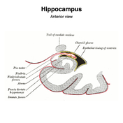
Hippocampus (Gray's illustration)
Published
27 Dec 2021
32% complete
Diagram
Case
Tela choroidea and choroid plexus of lateral ventricles (Gray's illustration)

Published
27 Dec 2021
44% complete
Diagram
Case
Internal capsule fibers (Gray's illustration)

Published
27 Dec 2021
32% complete
Diagram
Case
Fornix (Gray's illustration)

Published
27 Dec 2021
32% complete
Diagram
Case
Corpus striatum (Gray's illustration)

Published
27 Dec 2021
35% complete
Diagram
Case
Corona radiata (Gray's illustration)

Published
27 Dec 2021
32% complete
Diagram
Case
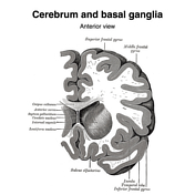
Basal ganglia (Gray's illustrations)
Published
27 Dec 2021
32% complete
Diagram
Case
Normal cardiac CT (volume render)

Published
23 Dec 2021
39% complete
CT
Case
Lateral talar process avulsion fracture

Published
20 Dec 2021
88% complete
X-ray
Case
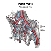
Pelvic veins (Gray's illustration)
Published
20 Dec 2021
32% complete
Diagram
Case
Superficial abdominal wall veins (Gray's illustration)

Published
20 Dec 2021
29% complete
Diagram
Case
Enchondroma with pathological fracture and incidental exostosis

Published
20 Dec 2021
75% complete
X-ray
Case
Neck veins (Gray's illustrations)

Published
20 Dec 2021
35% complete
Diagram
Case
Implantable contraceptive device

Published
20 Dec 2021
91% complete
X-ray
Case
Dural venous sinuses (Gray's illustrations)

Published
20 Dec 2021
32% complete
Diagram
Case
Veins of the scrotum (Gray's illustration)

Published
08 Dec 2021
35% complete
Diagram
Case
Female reproductive tract vessels (Gray's illustration)

Published
08 Dec 2021
35% complete
Diagram
Case
Superficial veins of the lower limb (Gray's illustration)
Published
08 Dec 2021
35% complete
Diagram
Case
Veins of the axilla (Gray's illustration)

Published
08 Dec 2021
35% complete
Diagram
Case
Tongue vessels (Gray's illustration)

Published
08 Dec 2021
35% complete
Diagram
Case
Vertebral venous plexuses (Gray's illustrations)

Published
08 Dec 2021
32% complete
Diagram
Case
Thyroid veins (Gray's illustration)

Published
08 Dec 2021
35% complete
Diagram
Case
Superficial veins of the elbow (Gray's illustration)

Published
08 Dec 2021
35% complete
Diagram
Case
Cavernous sinus (Gray's illustration)

Published
08 Dec 2021
32% complete
Diagram
Case
Superficial veins of the hand (Gray's illustration)

Published
08 Dec 2021
35% complete
Diagram
Case
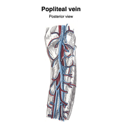
Popliteal vein (Gray's illustration)
Published
08 Dec 2021
32% complete
Diagram
Case
Orbital veins (Gray's illustration)
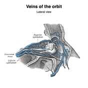
Published
08 Dec 2021
35% complete
Diagram
Case
Internal cerebral veins (Gray's illustration)

Published
07 Dec 2021
32% complete
Diagram
Case
CT angiogram head sagittal - labeling questions

Published
30 Nov 2021
40% complete
Annotated image
CT
Case
CT angiogram head coronal - labeling questions

Published
29 Nov 2021
40% complete
CT
Annotated image
Case
CT foot and ankle coronal - labeling questions

Published
25 Nov 2021
40% complete
CT
Annotated image
Case
CT foot and ankle axial - labeling questions

Published
23 Nov 2021
40% complete
CT
Annotated image
Case
CT foot and ankle sagittal - labeling questions

Published
22 Nov 2021
40% complete
CT
Annotated image
Case
Ischemic bowel due to internal hernia with perforation
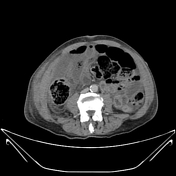
Published
21 Nov 2021
92% complete
CT
Case
Hereditary multiple exostosis

Published
21 Nov 2021
94% complete
X-ray
Case
CT wrist axial - labeling questions

Published
18 Nov 2021
40% complete
CT
Annotated image
Case
CT wrist sagittal - labeling questions

Published
17 Nov 2021
40% complete
CT
Annotated image
Case
CT wrist coronal - labeling questions

Published
17 Nov 2021
40% complete
Annotated image
CT
Case
MRI pituitary gland coronal T1 post contrast - labeling questions

Published
17 Nov 2021
40% complete
Annotated image
MRI
Case
MRI pituitary gland sagittal T1 post contrast - labeling questions

Published
16 Nov 2021
40% complete
MRI
Annotated image
Case
Romanus lesions - ankylosing spondylitis

Published
12 Nov 2021
80% complete
MRI
Case
CT temporal bone sagittal - labeling questions

Published
12 Nov 2021
40% complete
CT
Annotated image
Case
CT temporal bone coronal - labeling questions

Published
09 Nov 2021
40% complete
CT
Annotated image
Case
CT angiogram abdomen/pelvis sagittal - labeling questions

Published
08 Nov 2021
40% complete
Annotated image
CT
Case
Accessory PCA arising from the terminal ICA

Published
08 Nov 2021
79% complete
MRI
Case
Normal CTA abdomen and pelvis (female)

Published
08 Nov 2021
48% complete
CT
Case
Distal fibular and fifth metatarsal base fractures

Published
08 Nov 2021
88% complete
X-ray
Case
CT angiogram abdomen/pelvis coronal - labeling questions

Published
04 Nov 2021
40% complete
Annotated image
CT
Case
CT angiogram abdomen/pelvis axial - labeling questions

Published
03 Nov 2021
40% complete
CT
Annotated image
Case
MRI head axial T2 - labeling questions

Published
02 Nov 2021
40% complete
Annotated image
MRI
Case
MRI head sagittal T1 - labeling questions

Published
28 Oct 2021
40% complete
Annotated image
MRI
Case
Anal triangle (diagrams)

Published
27 Oct 2021
35% complete
Diagram
Case
Urogenital triangle (diagrams)

Published
27 Oct 2021
35% complete
Diagram
Case
CT cervical spine axial - labeling questions

Published
27 Oct 2021
40% complete
CT
Annotated image
Case
Female perineal muscles (Gray's illustration)

Published
26 Oct 2021
35% complete
Diagram
Case
Male perineal muscles (Gray's illustration)

Published
26 Oct 2021
35% complete
Diagram
Case
Male perineal fascia (Gray's illustration)

Published
26 Oct 2021
29% complete
Diagram
Case
Male pelvic and perineal fascia (Gray's illustrations)

Published
26 Oct 2021
29% complete
Diagram
Case
Levator ani (Gray's illustration)

Published
26 Oct 2021
32% complete
Diagram
Case
CT cervical spine coronal - labeling questions

Published
26 Oct 2021
40% complete
CT
Annotated image
Case
CT cervical spine sagittal - labeling questions

Published
20 Oct 2021
40% complete
CT
Annotated image
Case
Normal hip x-ray

Published
20 Oct 2021
35% complete
X-ray
Case
Clavicle x-ray - labeling questions

Published
19 Oct 2021
32% complete
Annotated image
Case
Fractured clavicle and bent ORIF cannulated screw

Published
05 Oct 2021
91% complete
X-ray
Case
Teeth anatomy - labeling questions
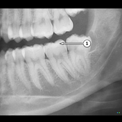
Published
22 Sep 2021
40% complete
X-ray
Annotated image
Case
OPG - labeling questions
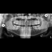
Published
22 Sep 2021
40% complete
X-ray
Annotated image
Case
Truncal venous development (Gray's illustrations)

Published
15 Sep 2021
35% complete
Diagram
Case
Hepatic venous development (Gray's illustration)

Published
15 Sep 2021
35% complete
Diagram
Case
Dural venous development (Gray's illustrations)

Published
15 Sep 2021
35% complete
Diagram
Case
Carotid artery development (Gray's illustration)
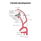
Published
15 Sep 2021
35% complete
Diagram
ADVERTISEMENT: Supporters see fewer/no ads


 Unable to process the form. Check for errors and try again.