


1,442 results found
Case
Normal hip x-ray

Published
20 Oct 2021
35% complete
X-ray
Case
Clavicle x-ray - labeling questions

Published
19 Oct 2021
32% complete
Annotated image
Case
Fractured clavicle and bent ORIF cannulated screw

Published
05 Oct 2021
91% complete
X-ray
Case
Teeth anatomy - labeling questions
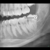
Published
22 Sep 2021
40% complete
X-ray
Annotated image
Case
OPG - labeling questions
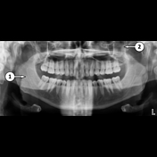
Published
22 Sep 2021
40% complete
X-ray
Annotated image
Case
Truncal venous development (Gray's illustrations)

Published
15 Sep 2021
35% complete
Diagram
Case
Hepatic venous development (Gray's illustration)

Published
15 Sep 2021
35% complete
Diagram
Case
Dural venous development (Gray's illustrations)

Published
15 Sep 2021
35% complete
Diagram
Case
Carotid artery development (Gray's illustration)
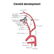
Published
15 Sep 2021
35% complete
Diagram
Case
Sinus venosus development (Gray's illustration)

Published
15 Sep 2021
29% complete
Diagram
Case
Aortic arches (Gray's illustration)

Published
14 Sep 2021
32% complete
Diagram
Case
Pneumothorax due to lymphangioleiomyomatosis
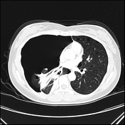
Published
01 Sep 2021
80% complete
X-ray
CT
Case
Superior ophthalmic vein thrombosis

Published
31 Aug 2021
92% complete
CT
Case
Cricothyroidotomy tube for severe epiglottitis

Published
31 Aug 2021
89% complete
CT
Case
Ankle and foot bone infarcts secondary to alcoholism

Published
31 Aug 2021
89% complete
X-ray
CT
Case
Femoral canal (Gray's illustrations)
Published
29 Aug 2021
35% complete
Diagram
Case
Femoral triangle and sheath (Gray's illustrations)

Published
29 Aug 2021
35% complete
Diagram
Case
Internal pudendal artery (Gray's illustrations)

Published
26 Aug 2021
32% complete
Diagram
Case
Subdural hemorrhage on CT perfusion

Published
26 Aug 2021
39% complete
CT
Case
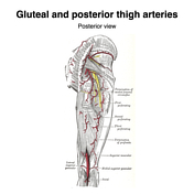
Gluteal arteries (Gray's illustration)
Published
26 Aug 2021
35% complete
Diagram
Case
Leg arteries (Gray's illustrations)

Published
26 Aug 2021
35% complete
Diagram
Case
Femoral artery (Gray's illustrations)

Published
26 Aug 2021
35% complete
Diagram
Case
Plantar arteries (Gray's illustrations)

Published
26 Aug 2021
32% complete
Diagram
Case
Left atrium and ventricle (Gray's illustration)

Published
26 Aug 2021
35% complete
Diagram
Case
Left and right ventricles (Gray's illustration)

Published
26 Aug 2021
35% complete
Diagram
Case
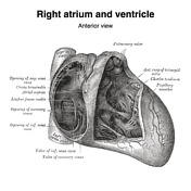
Right atrium and ventricle (Gray's illustration)
Published
26 Aug 2021
35% complete
Diagram
Case
Heart (Gray's illustrations)

Published
26 Aug 2021
32% complete
Diagram
Case
Genicular arteries (Gray's illustration)

Published
26 Aug 2021
32% complete
Diagram
Case
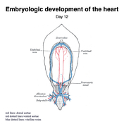
Embryologic development of the heart (Gray's illustrations)
Published
26 Aug 2021
32% complete
Diagram
Case
Atrioventricular bundle of His (Gray's illustration)

Published
26 Aug 2021
32% complete
Diagram
Case
Pulmonary arteries and veins (Gray's illustration)

Published
25 Aug 2021
35% complete
Diagram
Case
Visceral nerves of the thorax (Gray's illustration)

Published
25 Aug 2021
35% complete
Diagram
Case
Aortic valve opened up (Gray's illustration)

Published
25 Aug 2021
32% complete
Diagram
Case
Cardiac fibrous skeleton (Gray's illustration)

Published
25 Aug 2021
35% complete
Diagram
Case
Pericardial sinuses (Gray's illustration)

Published
25 Aug 2021
35% complete
Diagram
Case
Obturator internus (Gray's illustration)

Published
25 Aug 2021
32% complete
Diagram
Case
Gluteal and posterior thigh muscles (Gray's illustration)
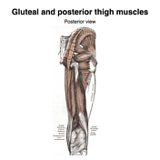
Published
25 Aug 2021
35% complete
Diagram
Case
Medial thigh muscles (Gray's illustration)

Published
25 Aug 2021
32% complete
Diagram
Case
Saphenous hiatus (Gray's illustration)

Published
25 Aug 2021
35% complete
Diagram
Case
Anterior thigh muscles (Gray's illustration)

Published
25 Aug 2021
32% complete
Diagram
Case
Posterior shoulder muscles (Gray's illustration)

Published
25 Aug 2021
32% complete
Diagram
Case
Sternoclavicular joint dislocation

Published
24 Aug 2021
92% complete
X-ray
CT
Case
Palmar aponeurosis (Gray's illustration)

Published
24 Aug 2021
35% complete
Diagram
Case
Thenar muscles (Gray's illustration)

Published
24 Aug 2021
35% complete
Diagram
Case
Intrinsic hand muscles (Gray's illustration)

Published
24 Aug 2021
35% complete
Diagram
Case
Hand interossei muscles (Gray's illustrations)

Published
24 Aug 2021
35% complete
Diagram
Case
Tendon sheaths of the wrist and hand (Gray's illustrations)

Published
24 Aug 2021
29% complete
Diagram
Case
Central line malpositioned in the aorta

Published
24 Aug 2021
86% complete
CT
X-ray
Case
Anterior process of calcaneum fracture

Published
24 Aug 2021
91% complete
X-ray
Case
Wrist cross section diagrams (Gray's illustrations)
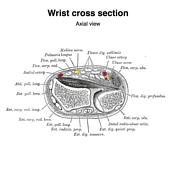
Published
18 Aug 2021
35% complete
Diagram
Case
Supinator (Gray's illustration)

Published
18 Aug 2021
32% complete
Diagram
Case
Posterior forearm muscles (Gray's illustrations)

Published
18 Aug 2021
32% complete
Diagram
Case
Anterior forearm muscles (Gray's illustrations)
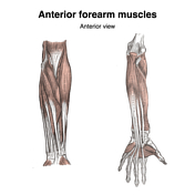
Published
18 Aug 2021
32% complete
Diagram
Case
Ulnocarpal synovial osteochondromatosis

Published
15 Aug 2021
82% complete
X-ray
Case
Posterior leg muscles (Gray's illustrations)
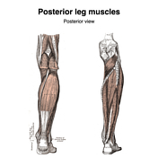
Published
15 Aug 2021
35% complete
Diagram
Case
Anterior and lateral leg muscles (Gray's illustration)

Published
15 Aug 2021
35% complete
Diagram
Case
Pectoral muscles (Gray's illustrations)

Published
15 Aug 2021
35% complete
Diagram
Case
Anterior abdominal wall (Gray's illustrations)

Published
15 Aug 2021
35% complete
Diagram
Case
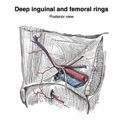
Inguinal canal and rings (Gray's illustrations)

Published
03 Aug 2021
32% complete
Diagram
Case
Ankle tendons (Gray's illustrations)

Published
03 Aug 2021
35% complete
Diagram
Case
Plantar foot muscles (Gray's illustrations)
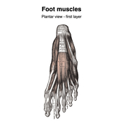
Published
03 Aug 2021
29% complete
Diagram
Case
Palatine bone (Gray's illustrations)

Published
03 Aug 2021
32% complete
Diagram
Case
Zygomatic bone (Gray's illustrations)

Published
03 Aug 2021
32% complete
Diagram
Case
Foot interossei muscles (Gray's illustrations)

Published
02 Aug 2021
35% complete
Diagram
Case
Rectus sheath (Gray's illustrations)

Published
02 Aug 2021
32% complete
Diagram
Case
Lateral process of talus fracture

Published
29 Jul 2021
79% complete
X-ray
Annotated image
Case
Essex-Lopresti fracture-disclocation

Published
29 Jul 2021
88% complete
X-ray
Case
Shoulder metastases

Published
22 Jul 2021
88% complete
X-ray
Case
Hallux sesamoid fracture

Published
22 Jul 2021
66% complete
X-ray
Case
Contrast filling the appendix post contrast swallow study

Published
22 Jul 2021
82% complete
X-ray
Case
Maxillary sinus (Gray's illustration)

Published
22 Jul 2021
35% complete
Diagram
Case
Hard palate (Gray's illustration)

Published
22 Jul 2021
35% complete
Diagram
Case
Dislocated reverse total shoulder arthroplasty

Published
13 Jul 2021
79% complete
X-ray
Case
Key foreign body on CXR

Published
13 Jul 2021
69% complete
X-ray
Case
Nasal conchae (Gray's illustration)

Published
02 Jul 2021
35% complete
Diagram
Case
Ethmoid bone (Gray's illustrations)

Published
02 Jul 2021
32% complete
Diagram
Case
Lacrimal bone (Gray's illustration)

Published
02 Jul 2021
32% complete
Diagram
Case
Maxilla (Gray's illustrations)

Published
02 Jul 2021
32% complete
Diagram
Case
Nasal and lacrimal bones (Gray's illustration)

Published
02 Jul 2021
35% complete
Diagram
Case
Nasal bone (Gray's illustrations)
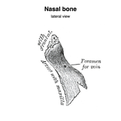
Published
02 Jul 2021
32% complete
Diagram
Case
Sphenoid bone (Gray's illustrations)
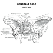
Published
01 Jul 2021
32% complete
Diagram
Case
Temporal bone (Gray's illustrations)
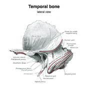
Published
23 Jun 2021
32% complete
Diagram
Case
Frontal bone (Gray's illustrations)

Published
23 Jun 2021
32% complete
Diagram
Case
Parietal bone (Gray's illustrations)

Published
23 Jun 2021
32% complete
Diagram
Case
Occipital bone (Gray's illustrations)

Published
23 Jun 2021
32% complete
Diagram
Case
Vomer (Gray's illustrations)

Published
22 Jun 2021
35% complete
Diagram
Case
Inferior nasal turbinate (Gray's illustrations)

Published
22 Jun 2021
32% complete
Diagram
Case
Normal spectral CTPA

Published
15 Jun 2021
62% complete
CT
Case
Periaortic abscess

Published
02 Jun 2021
91% complete
CT
Case
Avulsion of hallux toe nail

Published
02 Jun 2021
69% complete
X-ray
Case
Pathological fracture of the clavicle (multiple myeloma)

Published
02 Jun 2021
88% complete
X-ray
Case
Orbtial floor and ethmoidal sinuses (Gray's illustration)

Published
24 May 2021
35% complete
Diagram
Case
Medial wall of the orbit and maxillary sinus (Gray's illustration)
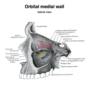
Published
24 May 2021
35% complete
Diagram
Case
Skull (Gray's illustrations)
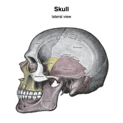
Published
18 May 2021
35% complete
Diagram
ADVERTISEMENT: Supporters see fewer/no ads


 Unable to process the form. Check for errors and try again.