


1,442 results found
Case
Cord presentation

Published
18 May 2021
85% complete
Ultrasound
Case
Deep anterior chest wall muscles (Gray's illustration)

Published
12 May 2021
32% complete
Diagram
Case
Deep back muscles (Gray's illustration)

Published
12 May 2021
35% complete
Diagram
Case
Deep neck muscles (Gray's illustration)

Published
12 May 2021
35% complete
Diagram
Case
Infratemporal fossa (Gray's illustration)

Published
12 May 2021
32% complete
Diagram
Case
Hyoid bone (Gray's illustration)

Published
05 May 2021
32% complete
Diagram
Case
Nasal cavity (Gray's illustrations)

Published
05 May 2021
35% complete
Diagram
Case
Pronator fat pad sign with a subtle distal radial fracture

Published
29 Apr 2021
91% complete
X-ray
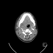
Case
Bipartite atlas

Published
24 Apr 2021
92% complete
CT
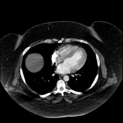
Case
Saddle pulmonary embolism with right ventricular strain and right atrial thrombus (spectral CT)

Published
24 Apr 2021
86% complete
CT
Case
Normal knee CT

Published
19 Apr 2021
83% complete
CT
Case
Cortical desmoid

Published
15 Apr 2021
84% complete
CT
X-ray
Case
Hangman fracture - Levine and Edwards type 2a

Published
13 Apr 2021
77% complete
CT
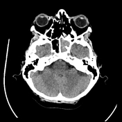
Case
Malignant otitis externa

Published
10 Apr 2021
71% complete
CT
Case
Pubic rami, acetabular and sacral fractures

Published
10 Apr 2021
95% complete
X-ray
CT
Case
Levine and Edwards classification of hangman fractures (diagrams)

Published
06 Apr 2021
35% complete
Diagram
Case
Pulmonary embolism on suboptimal CTPA (spectral low monoE)
Published
25 Mar 2021
95% complete
CT
Case
Segmental pulmonary embolism (spectral CTPA)

Published
25 Mar 2021
92% complete
CT
Case
Pulmonary embolism (spectral CTPA)

Published
25 Mar 2021
90% complete
CT
Case
Odontoid fracture (Anderson and D'Alonzo type 3, Roy-Camille type 2)

Published
25 Mar 2021
92% complete
CT
Case
Odontoid fracture (Anderson and D'Alonzo type 3, Roy-Camille type 1)

Published
25 Mar 2021
92% complete
CT
Case
Odontoid fracture (Anderson and D'Alonzo type 2, Roy-Camille type 3)

Published
25 Mar 2021
92% complete
CT
Case
Atlas (type 3b subtype 1) and axis (Anderson and D'Alonzo type 3, Roy-Camille type 2) fractures

Published
25 Mar 2021
95% complete
CT
MRI
Case
Infected ICA pseudoaneurysm
Published
24 Mar 2021
95% complete
CT
Case
Essex-Lopresti fracture-disclocation

Published
21 Mar 2021
88% complete
X-ray
Case
Occipital condyle fracture (type 1) and atlas transverse process fracture (type 5)

Published
17 Mar 2021
92% complete
CT
Case
Occipital condyle fracture (type 2) with extension into clivus

Published
17 Mar 2021
92% complete
CT
Case
Bilateral occipital condyle fracture (type 2)

Published
17 Mar 2021
92% complete
CT
Case
Occipital condyle fracture (type 3)

Published
17 Mar 2021
92% complete
CT
Case
Occipital condyle fracture (type 1)

Published
17 Mar 2021
92% complete
CT
Case
Bilateral occipital condyle fractures (type 3)

Published
17 Mar 2021
92% complete
CT
Case
Wrist CPPD

Published
15 Mar 2021
51% complete
X-ray
CT
Case
Atlanto-occipital dissociation (Traynelis type 1), C2 teardrop fracture, C6/7 facet joint dislocation

Published
15 Mar 2021
89% complete
CT
Case
Atlanto-occipital dissociation - Traynelis type 1

Published
15 Mar 2021
89% complete
CT
MRI
Case
Lester Jones tube
Published
15 Mar 2021
92% complete
CT
Case
Normal foot CT (with 3D VRTs)

Published
15 Mar 2021
42% complete
CT
Case
Gehweiler classification of atlas fractures (diagrams)

Published
11 Mar 2021
35% complete
Diagram
Case
Roy-Camille classification of C2 odontoid fractures (diagrams)

Published
03 Mar 2021
35% complete
Diagram
Case
Anderson and D'Alonzo classification of C2 odontoid fractures (diagrams)

Published
25 Feb 2021
35% complete
Diagram
Case
Muscles of mastication (Gray's illustration)
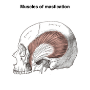
Published
24 Feb 2021
35% complete
Diagram
Case
Anderson and Montesano classification of occipital condyle fractures (diagrams)

Published
23 Feb 2021
35% complete
Diagram
Case
Traynelis classification of atlanto-occipital dissociation (diagrams)

Published
23 Feb 2021
35% complete
Diagram
Case
Talonavicular dislocation

Published
22 Feb 2021
91% complete
X-ray
Case
Subtle maxillary sinus fracture

Published
22 Feb 2021
89% complete
CT
Case
Incidental large fecolith

Published
22 Feb 2021
48% complete
X-ray
CT
Case
Fibroid red degeneration in pregnancy

Published
22 Feb 2021
75% complete
Ultrasound
Case
Delayed traumatic splenic pseudoaneurysms

Published
22 Feb 2021
83% complete
CT
DSA (angiography)
Case
Attempted bifid distal phalanx of the thumb

Published
22 Feb 2021
69% complete
X-ray
Case
Windswept knees

Published
22 Feb 2021
91% complete
X-ray
Case
Uterine version and flexion (diagrams)

Published
19 Feb 2021
22% complete
Diagram
Case
Thoracolumbar injury classification and severity score (table)

Published
16 Feb 2021
10% complete
Diagram
Case
Subaxial cervical spine injury classification (table)

Published
16 Feb 2021
10% complete
Diagram
Case
ECG gating (diagrams)

Published
09 Feb 2021
32% complete
Diagram
Case
Talar neck fracture - Hawkins type 3

Published
02 Feb 2021
87% complete
X-ray
CT
Case
Splenic injury - grade 5

Published
02 Feb 2021
92% complete
CT
Case
Medial malleolar and lateral talar dome fractures

Published
25 Jan 2021
78% complete
X-ray
CT
Case
Lymphatics of the thorax and abdomen (Gray's illustration)

Published
25 Jan 2021
29% complete
Diagram
Case
Pelvic lymphatics (Gray's illustrations)

Published
25 Jan 2021
35% complete
Diagram
Case
Lymphatics of the midgut (Gray's illustrations)

Published
25 Jan 2021
29% complete
Diagram
Case
Bell clapper (photo)

Published
21 Jan 2021
38% complete
Photo
Case
Bell clapper deformity (diagram)

Published
21 Jan 2021
35% complete
Diagram
Case
Salter Harris 4 fracture of index finger middle phalanx

Published
14 Jan 2021
85% complete
X-ray
Case
Lymphatics of the stomach and foregut (Gray's illustrations)

Published
14 Jan 2021
22% complete
Case
Lymphatics of the colon (Gray's illustration)

Published
11 Jan 2021
32% complete
Diagram
Case
Lymphatics of the bladder (Gray's illustration)

Published
11 Jan 2021
32% complete
Diagram
Case
Lymphatics of the prostate (Gray's illustration)

Published
11 Jan 2021
32% complete
Diagram
Case
Lymphatics of the uterus (Gray's illustration)

Published
11 Jan 2021
32% complete
Diagram
Case
Lymphatics of the tracheobronchial tree (Gray's illustration)

Published
30 Dec 2020
35% complete
Diagram
Case
Lymphatics of the lower limb (Gray's illustration)

Published
29 Dec 2020
35% complete
Diagram
Case
Lymphatics of the popliteal fossa (Gray's illustration)

Published
29 Dec 2020
35% complete
Diagram
Case
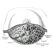
Female mammary gland (Gray's illustration)
Published
28 Dec 2020
32% complete
Diagram
Case
Lymphatics of the breast and axilla (Gray's illustration)

Published
28 Dec 2020
35% complete
Diagram
Case
Extensive rib plating for flail segment

Published
20 Dec 2020
94% complete
X-ray
Case
Ankle and foot interosseous ligaments (Gray's illustrations)

Published
20 Dec 2020
32% complete
Diagram
Case
Lymphatics of the upper limb (Gray's illustration)

Published
20 Dec 2020
35% complete
Diagram
Case
Lymphatics of the tongue (Gray's illustration)

Published
20 Dec 2020
32% complete
Diagram
Case
Lymphatics of the face (Gray's illustration)

Published
20 Dec 2020
29% complete
Diagram
Case
Lymphatics of the pharynx (Gray's illustration)

Published
20 Dec 2020
32% complete
Diagram
Case
Lymphatics of head and neck (Gray's illustration)

Published
17 Dec 2020
32% complete
Diagram
Case
Patellar tendon rupture

Published
17 Dec 2020
83% complete
X-ray
MRI
Case
Thoracic and right lymphatic ducts (Gray's illustration)

Published
17 Dec 2020
35% complete
Diagram
Case
Menstrual cup on trauma imaging

Published
17 Dec 2020
80% complete
X-ray
CT
Case
Hepatic abscess

Published
17 Dec 2020
89% complete
CT
Ultrasound
Case
Plantar ligaments of the foot (Gray's illustration)

Published
17 Dec 2020
35% complete
Diagram
Case
Lateral talocrural ligaments (Gray's illustration)

Published
17 Dec 2020
35% complete
Diagram
Case
Ankle and foot ligaments (Gray's illustrations)

Published
17 Dec 2020
35% complete
Diagram
Case
Subtalar ligaments (Gray's illustration)

Published
17 Dec 2020
35% complete
Diagram
Case
Patella (Gray's illustration)

Published
14 Dec 2020
32% complete
Diagram
Case
Knee joint capsule (Gray's illustrations)

Published
14 Dec 2020
35% complete
Diagram
Case
Hoffa's fat pad (Gray's illustration)

Published
14 Dec 2020
32% complete
Diagram
Case
Divergent Lisfranc injury

Published
13 Dec 2020
82% complete
X-ray
Case
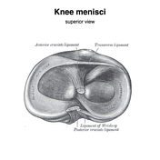
Knee menisci (Gray's illustrations)
Published
10 Dec 2020
35% complete
Diagram
Case
Internal knee ligaments (Gray's illustrations)

Published
10 Dec 2020
35% complete
Diagram
Case
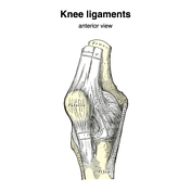
Knee ligaments (Gray's illustrations)
Published
10 Dec 2020
35% complete
Diagram
ADVERTISEMENT: Supporters see fewer/no ads


 Unable to process the form. Check for errors and try again.