


1,442 results found
Case
Food bolus (lateral neck x-ray)

Published
05 Oct 2020
91% complete
X-ray
Case
Temporomandibular joint anatomy (internal) (Gray's anatomy)

Published
05 Oct 2020
32% complete
Diagram
Case
Temporomandibular joint anatomy (medial) (Gray's anatomy)

Published
05 Oct 2020
35% complete
Diagram
Case
Temporomandibular joint anatomy (lateral) (Gray's anatomy)

Published
05 Oct 2020
32% complete
Diagram
Case
Typical thoracic vertebra (Gray's illustration)

Published
05 Oct 2020
35% complete
Diagram
Case
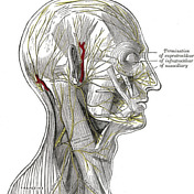
Trigeminal nerve cutaneous distribution (Gray's anatomy)

Published
24 Sep 2020
32% complete
Diagram
Case
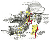
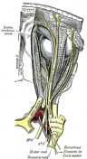
Mandibular division of the trigeminal nerve and submandibular and otic ganglia (Gray's illustration)
Published
20 Sep 2020
35% complete
Diagram
Case
Mandibular division of the trigeminal nerve (Gray's illustration)
Published
20 Sep 2020
35% complete
Diagram
Case
Elbow joint capsule (Gray's illustration)

Published
20 Sep 2020
32% complete
Diagram
Case
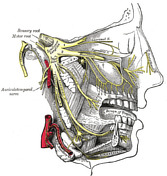
Pterygopalatine ganglion

Published
20 Sep 2020
35% complete
Diagram
Case
Maxillary division of the trigeminal nerve (Gray's illustration)

Published
20 Sep 2020
35% complete
Diagram
Case
Maxillary and mandibular divisions of the trigeminal nerve (Gray's illustration)
Published
20 Sep 2020
35% complete
Diagram
Case
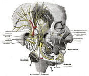
Nerves of the face, scalp and neck (Gray's illustration)
Published
20 Sep 2020
35% complete
Diagram
Case
Anatomy of the genicular ganglion (Gray's illustration)

Published
19 Sep 2020
35% complete
Diagram
Case
Nerves of the orbit (Gray's illustration)

Published
19 Sep 2020
35% complete
Diagram
Case
Anatomy of the ophthalmic division of the trigeminal nerve (Gray's illustration)
Published
19 Sep 2020
35% complete
Diagram
Case
Anatomy of the oculomotor nerve (Gray's illustration)

Published
19 Sep 2020
35% complete
Diagram
Case
Cranial nerve nuclei (axial diagrams)

Published
18 Sep 2020
25% complete
Diagram
Case
Pontine anatomy - CN V (diagram)

Published
18 Sep 2020
32% complete
Diagram
Case
Olfactory nerve (Gray's illustration)

Published
17 Sep 2020
35% complete
Diagram
Case
Optic nerve and chiasm (Gray's illustration)

Published
17 Sep 2020
35% complete
Diagram
Case
Medial elbow ligaments (Gray's illustration)

Published
16 Sep 2020
32% complete
Diagram
Case
Lateral elbow ligaments (Gray's illustration)

Published
16 Sep 2020
35% complete
Diagram
Case
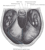
Cranial meninges and falx (Gray's illustration)

Published
14 Sep 2020
35% complete
Diagram
Case
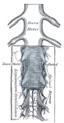
Spinal meninges (Gray's illustration)
Published
14 Sep 2020
35% complete
Diagram
Case
Tentorium cerebelli (Gray's illustration)
Published
14 Sep 2020
32% complete
Diagram
Case
Insular cortex (Gray's illustration)

Published
14 Sep 2020
32% complete
Diagram
Case
Basal ganglia (Gray's illustration)
Published
14 Sep 2020
35% complete
Diagram
Case
Heel soft tissue infection with gas forming organism

Published
13 Sep 2020
88% complete
X-ray
Case
Brainstem cross-sectional anatomy (diagrams)

Published
08 Sep 2020
29% complete
Diagram
Case
Optic radiations (Gray's illustration)

Published
08 Sep 2020
35% complete
Diagram
Case
Descending spinal tracts (Gray's illustration)

Published
08 Sep 2020
35% complete
Diagram
Case
Ascending spinal tracts (Gray's illustration)

Published
08 Sep 2020
35% complete
Diagram
Case
Cranial nerves (Gray's illustration)

Published
08 Sep 2020
32% complete
Diagram
Case
Spinal cord (Gray's illustration)

Published
07 Sep 2020
35% complete
Diagram
Case
Cauda equina (Gray's illustration)

Published
07 Sep 2020
35% complete
Diagram
Case
Pharyngeal constrictors (Gray's illustration)

Published
07 Sep 2020
35% complete
Diagram
Case
Forearm deep arterial anatomy (Gray's illustration)

Published
06 Sep 2020
35% complete
Diagram
Case
Anterior thorax and abdominal wall arterial supply (Gray's illustration)
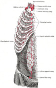
Published
06 Sep 2020
35% complete
Diagram
Case
External carotid artery (Gray's illustration)

Published
06 Sep 2020
32% complete
Diagram
Case
Scapular arterial anastomoses (Gray's illustration)

Published
06 Sep 2020
32% complete
Diagram
Case
Circle of Willis (Gray's illustration)

Published
06 Sep 2020
32% complete
Diagram
Case
Inferior mesenteric artery (Gray's illustration)

Published
06 Sep 2020
35% complete
Diagram
Case
Spinal cord cross section (Gray's illustration)

Published
06 Sep 2020
35% complete
Diagram
Case
Cranial nerves in the posterior fossa (Gray's illustration)

Published
06 Sep 2020
35% complete
Diagram
Case
Brainstem tracts (Gray's illustrations)

Published
06 Sep 2020
32% complete
Diagram
Case
Pyramids and olives (Gray's illustration)
Published
03 Sep 2020
35% complete
Diagram
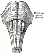
Case
Cerebellar peduncles (Gray's illustration)

Published
03 Sep 2020
35% complete
Diagram
Case
Colliculi connections (Gray's illustration)

Published
03 Sep 2020
35% complete
Diagram
Case
Interconnection of cranial nerve nuclei (Gray's illustration)

Published
03 Sep 2020
35% complete
Diagram
Case
Cerebellum inferior surface (Gray's illustration)

Published
03 Sep 2020
35% complete
Diagram
Case
Cerebellum superior surface (Gray's illustration)
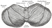
Published
03 Sep 2020
32% complete
Diagram
Case
Cerebellar peduncles (Gray's illustration)

Published
03 Sep 2020
35% complete
Diagram
Case
Dentate nucleus (Gray's illustration)

Published
03 Sep 2020
32% complete
Diagram
Case
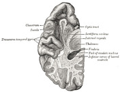
Anatomy of the lateral ventricles (Gray's illustration)

Published
03 Sep 2020
32% complete
Diagram
Case
Upper-level pons (Gray's illustration)

Published
03 Sep 2020
32% complete
Diagram
Case
Mid-level medulla anatomy (Gray's illustration)

Published
03 Sep 2020
32% complete
Diagram
Case
Decussation of fibers in the brainstem (Gray's illustration)

Published
03 Sep 2020
35% complete
Diagram
Case
Blood supply of the orbit (Gray's illustration)

Published
02 Sep 2020
35% complete
Diagram
Case
Surgical triangles of the neck (Gray's illustration)

Published
02 Sep 2020
35% complete
Diagram
Case
Anatomy of the penis (Gray's illustration)

Published
02 Sep 2020
35% complete
Diagram
Case
Venous drainage of the penis (Gray's illustration)

Published
02 Sep 2020
35% complete
Diagram
Case
Arterial supply of the penis (Gray's illustration)

Published
02 Sep 2020
35% complete
Diagram
Case
Testes and epididymis (Gray's illustration)

Published
02 Sep 2020
35% complete
Diagram
Case
Testes and spermatic cord (Gray's illustration)

Published
02 Sep 2020
35% complete
Diagram
Case
Prostate gland and seminal vesicles (Gray's illustration)

Published
02 Sep 2020
35% complete
Diagram
Case
Upper medulla anatomy - CN X (diagram)
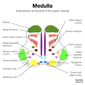
Published
02 Sep 2020
22% complete
Diagram
Case
Scrotal ring

Published
01 Sep 2020
50% complete
X-ray
Case
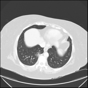
Esophageal rupture post stricture dilatation

Published
01 Sep 2020
65% complete
CT
Case
Normal breast MRI - dense breasts

Published
01 Sep 2020
86% complete
MRI
Case
Upper medulla anatomy - CN IX (diagram)

Published
01 Sep 2020
32% complete
Diagram
Case
Upper medulla anatomy - CN VIII (diagram)

Published
01 Sep 2020
32% complete
Diagram
Case
Finger PIP joint septic arthritis and osteomyelitis

Published
01 Sep 2020
94% complete
X-ray
Case
Toxic megacolon

Published
01 Sep 2020
95% complete
X-ray
CT
Case
Osteophyte-induced atelectasis and fibrosis
Published
01 Sep 2020
86% complete
CT
Case
Swallowed dentures (chest x-ray)

Published
01 Sep 2020
88% complete
X-ray
Case
Dislocated first carpometacarpal joint

Published
25 Aug 2020
79% complete
X-ray
Case
Glenoid version measurement: scapular blade method (diagram)

Published
19 Aug 2020
44% complete
Annotated image
Case
Lower pons anatomy - CN VII (diagram)

Published
18 Aug 2020
22% complete
Diagram
Case
Lower pons anatomy - CN VI (diagram)

Published
18 Aug 2020
22% complete
Diagram
Case
Upper medulla anatomy - CN XII (diagram)

Published
17 Aug 2020
32% complete
Diagram
Case
Upper medulla anatomy - CN XI (diagram)

Published
17 Aug 2020
32% complete
Diagram
Case
Subclavian artery traumatic injury

Published
07 Aug 2020
92% complete
CT
X-ray
DSA (angiography)
Case
Renal artery stenosis on captopril renal MAG3 nuclear study
Published
30 Jul 2020
94% complete
Nuclear medicine
Case
Normal captopril renal MAG3 nuclear study

Published
29 Jul 2020
94% complete
Nuclear medicine
Case
Haemophagocytosis

Published
29 Jul 2020
77% complete
CT
Case
Normal common hepatic artery angiogram

Published
29 Jul 2020
29% complete
DSA (angiography)
Case
Normal inferior mesenteric artery angiogram

Published
29 Jul 2020
57% complete
DSA (angiography)
Case
Dilated esophagus due to tight gastric band

Published
28 Jul 2020
94% complete
X-ray
Case
Normal renal MAG3 nuclear study

Published
27 Jul 2020
94% complete
Nuclear medicine
Case
Ligamentum teres avulsion fracture of femoral head

Published
24 Jul 2020
89% complete
X-ray
CT
Case
Fracture of the anterior process of the calcaneum on lateral ankle radiograph

Published
24 Jul 2020
88% complete
X-ray
Case
Normal breast MRI (fatty breasts)

Published
23 Jul 2020
77% complete
MRI
Case
Malignant pleural effusion

Published
23 Jul 2020
92% complete
X-ray
CT
ADVERTISEMENT: Supporters see fewer/no ads


 Unable to process the form. Check for errors and try again.