
Patella alta
Updates to Article Attributes
Patella alta, or a high riding-riding patella, describes a situation where the position of the patella is considered high. It may be idiopathic or may result secondary to a patellar tendon rupture.
Epidemiology
Associations
Several conditions are known to be associated with patella alta, including:
idiopathic retropatellar pain 5
recurrent dislocation of the patella 2
knee joint effusion 2
Patella alta may also occur as a result of spastic cerebral palsy 6.
Radiographic features
On sagittal images, it is usually considered when the Insall-Salvati ratio is greater than 1.3-1.5 1-3.
On radiography, it is important that standard positioning is used, with the knee flexed at 30 degrees. An off-angle x-ray beam or non-standard positioning may result in a spuriously abnormal ratio.
-<p><strong>Patella alta</strong>, or a <strong>high riding patella</strong>, describes a situation where the position of the patella is considered high. It may be idiopathic or may result secondary to a <a href="/articles/patellar-tendon-rupture">patellar tendon rupture</a>. </p><h4>Epidemiology</h4><h5>Associations</h5><p>Several conditions are known to be associated with patella alta, including:</p><ul>-<li>idiopathic retropatellar pain <sup>5</sup>-</li>-<li>recurrent dislocation of the patella <sup>2</sup>-</li>-<li>-<a href="/articles/chondromalacia-patellae">chondromalacia patellae</a> <sup>2</sup>-</li>-<li>knee joint effusion <sup>2</sup>-</li>- +<p><strong>Patella alta</strong>, or a <strong>high-riding patella</strong>, describes a situation where the position of the patella is considered high. It may be idiopathic or may result secondary to a <a href="/articles/patellar-tendon-rupture">patellar tendon rupture</a>. </p><h4>Epidemiology</h4><h5>Associations</h5><p>Several conditions are known to be associated with patella alta, including:</p><ul>
- +<li><p>idiopathic retropatellar pain <sup>5</sup></p></li>
- +<li><p>recurrent dislocation of the patella <sup>2</sup></p></li>
- +<li><p><a href="/articles/chondromalacia-patellae">chondromalacia patellae</a> <sup>2</sup></p></li>
- +<li><p>knee joint effusion <sup>2</sup></p></li>
References changed:
- 1. Shabshin N, Schweitzer M, Morrison W, Parker L. MRI Criteria for Patella Alta and Baja. Skeletal Radiol. 2004;33(8):445-50. <a href="https://doi.org/10.1007/s00256-004-0794-6">doi:10.1007/s00256-004-0794-6</a> - <a href="https://www.ncbi.nlm.nih.gov/pubmed/15221214">Pubmed</a>
- 2. Ali S, Helmer R, Terk M. Patella Alta: Lack of Correlation Between Patellotrochlear Cartilage Congruence and Commonly Used Patellar Height Ratios. AJR Am J Roentgenol. 2009;193(5):1361-6. <a href="https://doi.org/10.2214/AJR.09.2729">doi:10.2214/AJR.09.2729</a> - <a href="https://www.ncbi.nlm.nih.gov/pubmed/19843754">Pubmed</a>
- 3. Insall J & Salvati E. Patella Position in the Normal Knee Joint. Radiology. 1971;101(1):101-4. <a href="https://doi.org/10.1148/101.1.101">doi:10.1148/101.1.101</a> - <a href="https://www.ncbi.nlm.nih.gov/pubmed/5111961">Pubmed</a>
- 4. Miller T, Staron R, Feldman F. Patellar Height on Sagittal MR Imaging of the Knee. AJR Am J Roentgenol. 1996;167(2):339-41. <a href="https://doi.org/10.2214/ajr.167.2.8686598">doi:10.2214/ajr.167.2.8686598</a> - <a href="https://www.ncbi.nlm.nih.gov/pubmed/8686598">Pubmed</a>
- 5. Kannus P. Long Patellar Tendon: Radiographic Sign of Patellofemoral Pain Syndrome--A Prospective Study. Radiology. 1992;185(3):859-63. <a href="https://doi.org/10.1148/radiology.185.3.1438776">doi:10.1148/radiology.185.3.1438776</a> - <a href="https://www.ncbi.nlm.nih.gov/pubmed/1438776">Pubmed</a>
- 6. Lotman D. Knee Flexion Deformity and Patella Alta in Spastic Cerebral Palsy. Dev Med Child Neurol. 1976;18(3):315-9. <a href="https://doi.org/10.1111/j.1469-8749.1976.tb03653.x">doi:10.1111/j.1469-8749.1976.tb03653.x</a> - <a href="https://www.ncbi.nlm.nih.gov/pubmed/939346">Pubmed</a>
- 1. Shabshin N, Schweitzer ME, Morrison WB et-al. MRI criteria for patella alta and baja. Skeletal Radiol. 2004;33 (8): 445-50. <a href="http://dx.doi.org/10.1007/s00256-004-0794-6">doi:10.1007/s00256-004-0794-6</a> - <a href="http://www.ncbi.nlm.nih.gov/pubmed/15221214">Pubmed citation</a><div class="ref_v2"></div>
- 2. Ali SA, Helmer R, Terk MR. Patella alta: lack of correlation between patellotrochlear cartilage congruence and commonly used patellar height ratios. AJR Am J Roentgenol. 2009;193 (5): 1361-6. <a href="http://dx.doi.org/10.2214/AJR.09.2729">doi:10.2214/AJR.09.2729</a> - <a href="http://www.ncbi.nlm.nih.gov/pubmed/19843754">Pubmed citation</a><div class="ref_v2"></div>
- 3. Insall, John; Salvati, Eduardo. Radiology. <a href="http://dx.doi.org/10.1148/101.1.101">doi:10.1148/101.1.101</a><div class="ref_v2"></div>
- 4. Miller TT, Staron RB, Feldman F. Patellar height on sagittal MR imaging of the knee. AJR Am J Roentgenol. 1996;167 (2): 339-41. <a href="http://www.ajronline.org/cgi/content/abstract/167/2/339">AJR Am J Roentgenol (abstract)</a> - <a href="http://www.ncbi.nlm.nih.gov/pubmed/8686598">Pubmed citation</a><div class="ref_v2"></div>
- 5. Kannus PA. Long patellar tendon: radiographic sign of patellofemoral pain syndrome--a prospective study. Radiology. 1992;185 (3): 859-63. <a href="http://radiology.rsna.org/content/185/3/859.abstract">Radiology (abstract)</a> - <a href="http://www.ncbi.nlm.nih.gov/pubmed/1438776">Pubmed citation</a><div class="ref_v2"></div>
- 6. Lotman DB. Knee flexion deformity and patella alta in spastic cerebral palsy. Dev Med Child Neurol. 1976;18 (3): 315-9. <a href="http://www.ncbi.nlm.nih.gov/pubmed/939346">Pubmed citation</a><span class="auto"></span>
Image 3 X-ray (Lateral) ( destroy )

Image 3 X-ray (Lateral) ( update )

Image 4 X-ray (Lateral) ( update )
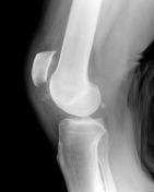
Image 5 X-ray (Lateral) ( update )

Image 6 MRI (PD) ( update )

Image 7 MRI (T1) ( update )

Image 8 MRI (PD) ( update )

Image 9 X-ray (Lateral) ( create )


 Unable to process the form. Check for errors and try again.