
Patient Data
Note: This case has been tagged as "legacy" as it no longer meets image preparation and/or other case publication guidelines.


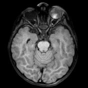
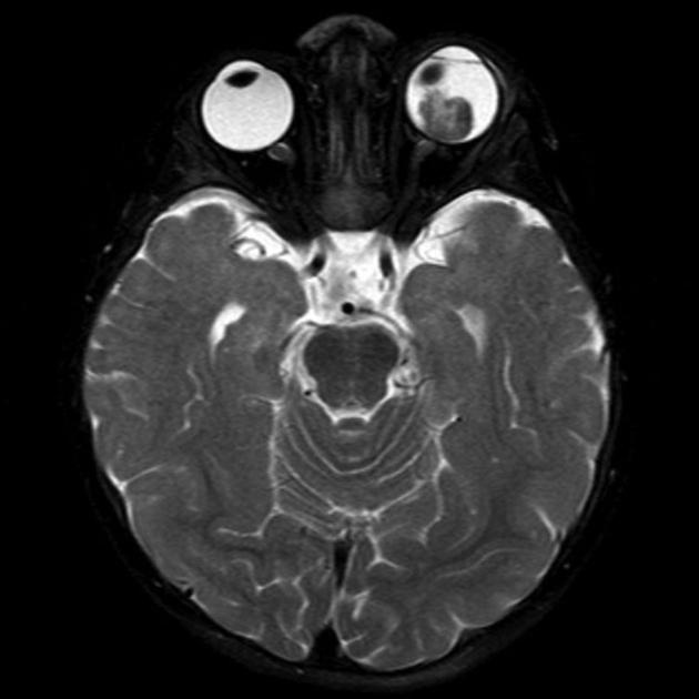
Selected MRI images through the orbits. On the left there is a dominant mass of decreased signal intensity on the T2 weighted images and mild contrast enhancement on the T1 weighted images. This is associated with dislocation of the lens and increased signal intensity within the globe on the T1 weighted sequences suggesting subacute hemorrhage.
On the right, there are two mass lesions identified best seen on coronal images. The largest is identified in the inferior medial quadrant of the right globe and the second separate lesion is identified superiorly in the midline posteriorly. Both lesions are of decreased signal intensity on the T2 weighted images.
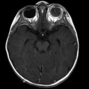
Note the phase mismapping artefact on the post contrast T1 sequence resulting in an apparent area of contrast enhancement peripherally in the left occipital lobe.
Case Discussion
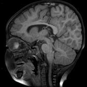
MRI of a child demonstrates bilateral retinoblastomas.


 Unable to process the form. Check for errors and try again.