Presentation
Cough
Patient Data

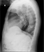
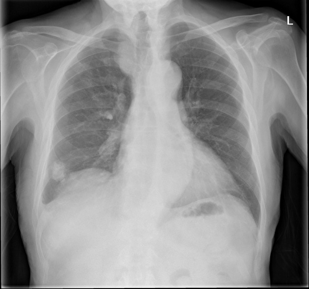
Calcified mass projected over the right lower lobe
Prominent right paratracheal mass.
The heart size is normal. No focal consolidation. Small right pleural effusion.
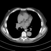

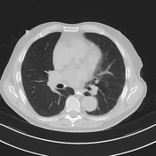

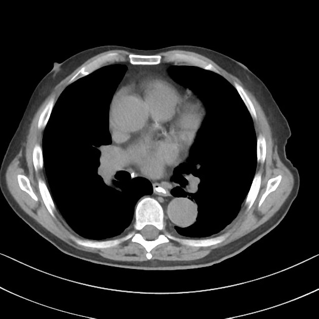
Calcified mass seen on CXR is actually a pleural plaque. There are two other calcified foci within the thorax: more inferiorly along the right pleura and involving the posterior mediastinal pleura.
Ectatic brachiocephalic trunk and right subclavian artery to the right of the trachea, this accounts for right paratracheal 'mass' identified on CXR.
Case Discussion
The density of this lesion on CXR should suggest a calcified lesion. In assessing a lung mass, ask whether this could be pleural, chest wall or elsewhere.


 Unable to process the form. Check for errors and try again.