
Presentation
CT staging after colonoscopy and ileocecal biopsy.
Patient Data
Age: 75 years
Gender: Female


Download
Info

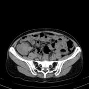
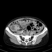
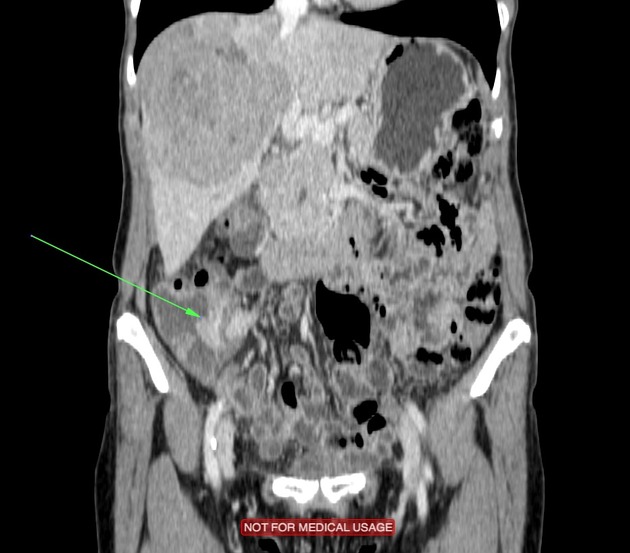
Large lesions in the right hepatic lobe, predominantly hypervascular with central areas of necrosis. Hypervascular solid lesions involving internal intestinal walls in the region of the ileocecal valve.
From the case:
Terminal ileum carcinoid tumor with hepatic metastasis
Download
Info
Note that the cecal wall is surrounded by two sides.
Case Discussion
The histopathological diagnosis confirmed the carcinoid tumor.
Carcinoid tumors are neuroendocrine neoplasms of diffuse cellular system, and nowadays the term neuroendocrine tumor has been proposed for this entity 1.
Diagnosis is made at the mean age of 50 years. The most frequent locations of carcinoid tumors are the gastrointestinal tract (73.7%) and respiratory system (25.1%) 1.


 Unable to process the form. Check for errors and try again.