Citation, DOI, disclosures and article data
Citation:
Deng F, Absent posterior limb sign. Reference article, Radiopaedia.org (Accessed on 27 Mar 2025) https://doi.org/10.53347/rID-84508
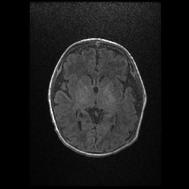
The absent posterior limb sign is one of the main MRI findings of prognostic significance in term neonates with suspected hypoxic-ischemic brain injury. An absent posterior limb sign is defined as loss of the normally distinct hyperintensity on T1-weighted images in the posterior part of the posterior limb of the internal capsule in neonates born at term (after 37 weeks gestation) 1-4. The signal intensity of the posterior limb of internal capsule can be compared to that of the posterolateral putamen, which is normally less intense than the posterior third to half of the posterior limb of internal capsule due to the latter's myelination; this relationship is inverted in hypoxic-ischemic injury 2,5. Correlation with T2-weighted images can help localize the posterior limb of internal capsule and show loss of the normal T2 hypointense focus in the same locale 4.
The absent posterior limb sign is associated with the basal ganglia-thalamus pattern of severe hypoxic-ischemic injury. It portends an adverse developmental outcome or death, while its absence (ie, normal hyperintensity of the posterior limb) suggests a favorable developmental outcome later in childhood 1,2.
The timing of imaging is important for the predictive performance of this sign. The posterior limb may appear falsely normal even in neonates with severe brain injury in the first few days of life, so imaging should be conducted after 3 days 1,3,6. Myelination of the posterior limb of internal capsule occurs around 36 weeks gestation, so this sign cannot be applied in the same time frame to neonates born preterm (before 37 weeks gestation) 4. Imaging often occurs after a period of therapeutic hypothermia, but this intervention does not negate the prognostic value of abnormal findings like the absent posterior limb sign 6.
-
1. Rutherford MA, Pennock JM, Counsell SJ, Mercuri E, Cowan FM, Dubowitz LM, Edwards AD. Abnormal magnetic resonance signal in the internal capsule predicts poor neurodevelopmental outcome in infants with hypoxic-ischemic encephalopathy. (1998) Pediatrics. 102 (2 Pt 1): 323-8. doi:10.1542/peds.102.2.323 - Pubmed
-
2. Liauw L, van der Grond J, van den Berg-Huysmans AA, Laan LA, van Buchem MA, van Wezel-Meijler G. Is there a way to predict outcome in (near) term neonates with hypoxic-ischemic encephalopathy based on MR imaging?. (2008) AJNR. American journal of neuroradiology. 29 (9): 1789-94. doi:10.3174/ajnr.A1188 - Pubmed
-
3. Ghei SK, Zan E, Nathan JE, Choudhri A, Tekes A, Huisman TA, Izbudak I. MR imaging of hypoxic-ischemic injury in term neonates: pearls and pitfalls. (2014) Radiographics : a review publication of the Radiological Society of North America, Inc. 34 (4): 1047-61. doi:10.1148/rg.344130080 - Pubmed
-
4. Heinz ER, Provenzale JM. Imaging findings in neonatal hypoxia: a practical review. (2009) AJR. American journal of roentgenology. 192 (1): 41-7. doi:10.2214/ajr.08.1321 - Pubmed
-
5. Liauw L, Palm-Meinders IH, van der Grond J, Leijser LM, le Cessie S, Laan LA, Heeres BC, van Buchem MA, van Wezel-Meijler G. Differentiating normal myelination from hypoxic-ischemic encephalopathy on T1-weighted MR Images: a new approach. (2007) AJNR. American journal of neuroradiology. 28 (4): 660-5. Pubmed
-
6. Cheong JL, Coleman L, Hunt RW, Lee KJ, Doyle LW, Inder TE, Jacobs SE. Prognostic utility of magnetic resonance imaging in neonatal hypoxic-ischemic encephalopathy: substudy of a randomized trial. (2012) Archives of pediatrics & adolescent medicine. 166 (7): 634-40. doi:10.1001/archpediatrics.2012.284 - Pubmed
Promoted articles (advertising)


 Unable to process the form. Check for errors and try again.