
Accessory muscles of the ankle
Citation, DOI, disclosures and article data
At the time the article was created Joachim Feger had no recorded disclosures.
View Joachim Feger's current disclosuresAt the time the article was last revised Joachim Feger had no recorded disclosures.
View Joachim Feger's current disclosures- Ankle accessory muscles
- Accessory muscles (ankle)
- Accessory muscle of the ankle
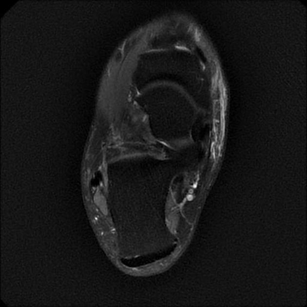
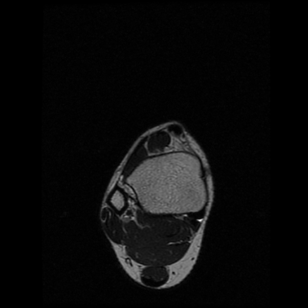
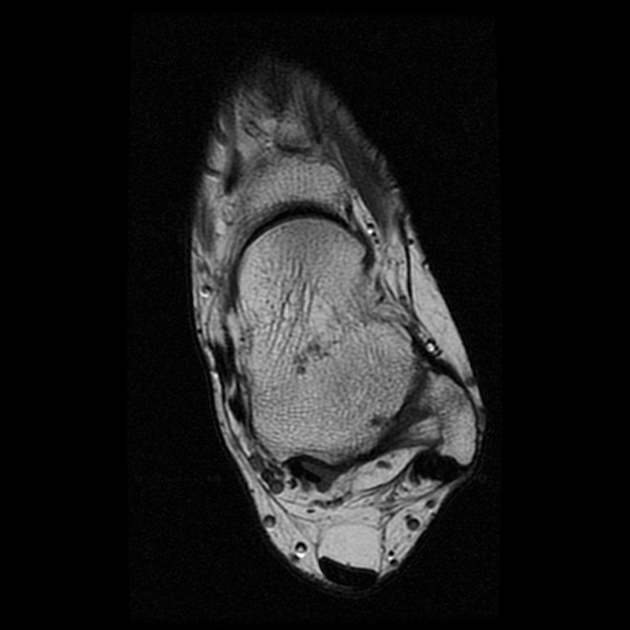
Accessory muscles of the ankle are muscular anatomical variants that are usually asymptomatic but rarely cause symptoms or might be encountered on imaging studies.
The following accessory muscles around the ankle have been described 1-4:
- posteromedial/flexor compartment
- accessory flexor digitorum longus muscle (deep to the flexor retinaculum)
- accessory soleus muscle (superficial to the flexor retinaculum)
- peroneocalcaneus internus muscle (rare, deep to the flexor retinaculum)
- tibiocalcaneus internus muscle (unknown, deep to the flexor retinaculum)
- lateral/peroneal compartment
-
peroneus quartus muscles
- peroneocalcaneus externus muscle
- peroneus digiti minimi muscle
- peroneus accessorius muscle
-
peroneus quartus muscles
- anterior/extensor compartment
On this page:
Radiographic features
Accessory muscles look like normal muscles in an abnormal location on imaging studies.
Radiology report
The radiological report should include a description of the following features:
- accessory muscle with location
- signs of impingement
- nerve compression syndromes
Clinical importance
Accessory muscles might mimic pathological conditions such as tumours or mass lesions.
Related pathology
Accessory muscles of the ankle have been implicated in the following clinical conditions 3,4:
- ankle impingement syndromes
- tarsal tunnel syndrome
- chronic ankle pain
- hindfoot deformity
References
- 1. Aparisi Gómez M, Aparisi F, Bartoloni A et al. Anatomical Variation in the Ankle and Foot: From Incidental Finding to Inductor of Pathology. Part I: Ankle and Hindfoot. Insights Imaging. 2019;10(1):74. doi:10.1186/s13244-019-0746-2 - Pubmed
- 2. Sookur P, Naraghi A, Bleakney R, Jalan R, Chan O, White L. Accessory Muscles: Anatomy, Symptoms, and Radiologic Evaluation. Radiographics. 2008;28(2):481-99. doi:10.1148/rg.282075064 - Pubmed
- 3. Cheung Y. Normal Variants. Magn Reson Imaging Clin N Am. 2017;25(1):11-26. doi:10.1016/j.mric.2016.08.002 - Pubmed
- 4. Carroll JF. Accessory Muscles of the Ankle. Radsource – November 2008. MRI Web Clinic
Incoming Links
Related articles: Anatomy: Lower limb
- skeleton of the lower limb
- joints of the lower limb
-
hip joint
- ligaments
- muscles
- additional structures
- hip joint capsule
- zona orbicularis
- iliotibial band
-
hip bursae
- anterior
- iliopsoas bursa (iliopectineal bursa)
- lateral
- subgluteal bursae
- greater trochanteric bursa (subgluteus maximus bursa)
- subgluteus medius bursa
- subgluteus minimus bursa
- gluteofemoral bursa
- subgluteal bursae
- postero-inferior
- anterior
- ossification centres
-
knee joint
- ligaments
- anterior cruciate ligament
- posterior cruciate ligament
- medial collateral ligament
- lateral collateral ligament
- meniscofemoral ligament (mnemonic)
-
posterolateral ligamentous complex
- arcuate ligament
- patellar tendon and quadriceps tendon
- anterolateral ligament
- posterior oblique ligament
- oblique popliteal ligament
- medial patellofemoral ligament
- additional structures
- extensor mechanism of the knee
- groove for the popliteus tendon
- knee bursae
- anterior bursae
- medial bursae
- lateral bursae
- posterior bursae
- knee capsule
- lateral patellar retinaculum
- medial patellar retinaculum
- menisci
- pes anserinus (mnemonic)
- ossification centres
- ligaments
- tibiofibular joints
-
ankle joint
- regional anatomy
- medial ankle
- lateral ankle
- anterior ankle
- ligaments
- medial collateral (deltoid) ligament
- lateral collateral ligament
- additional structures
- ankle bursae
- ossification centres of the ankle
- variants
- regional anatomy
- foot joints
- subtalar joint
- mid-tarsal (Chopart) joint
-
tarsometatarsal (Lisfranc) joint
- ligaments
- intermetatarsal joint
- metatarsophalangeal joint
- interphalangeal joint
- ossification centres
-
hip joint
- spaces of the lower limb
-
muscles of the lower limb
- muscles of the pelvic group
- muscles of the thigh
- muscles of the leg
- anterior compartment of the leg
- posterior compartments of the leg
- lateral compartment of the leg
- muscles of the foot
- dorsal muscles
- plantar muscles
- 1st layer
- 2nd layer
- 3rd layer
- 4th layer
- accessory muscles of the lower limb
- accessory gluteal muscles
-
accessory muscles of the ankle
- accessory peroneal muscles
- accessory flexor digitorum longus muscle
- accessory soleus muscle
- peroneocalcaneus internus muscle
- tibiocalcaneus internus muscle
- extensor hallucis capsularis tendon
- anterior fibulocalcaneus muscle
- accessory extensor digiti secundus muscle
- tibioastragalus anticus of Gruber muscle
- vascular supply of the lower limb
- arterial supply of the lower limb
- venous drainage of the lower limb
- innervation of the lower limb
- lymphatic system of the lower limb
- lymphatic pathways
- anteromedial group
- anterolateral group
- posteromedial group
- posterolateral group
- lower limb lymph nodes
- lymphatic pathways


 Unable to process the form. Check for errors and try again.