Acromion fracture
Citation, DOI, disclosures and article data
At the time the article was created Bahman Rasuli had no recorded disclosures.
View Bahman Rasuli's current disclosuresAt the time the article was last revised Frank Gaillard had no financial relationships to ineligible companies to disclose.
View Frank Gaillard's current disclosuresThe acromion process is the lateral projection of the scapula spine that extends anteriorly. Fractures of the scapula are uncommon injuries and account for ~3% of all shoulder fractures 1,2 while isolated acromion fractures occur rarely and account for only 9% of all scapular fractures 3.
On this page:
Pathology
Mechanisms of injury
Acromial fractures usually occur as the result of direct trauma to the shoulder or superior dislocation of the humeral head 4.
Radiographic features
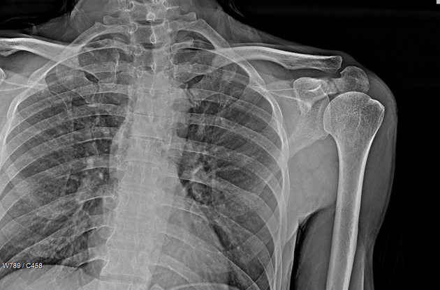
Shoulder radiographs are the first diagnostic investigation and the lateral scapula shoulder or Y view is an excellent projection to evaluate the coracoid and acromion process 5.
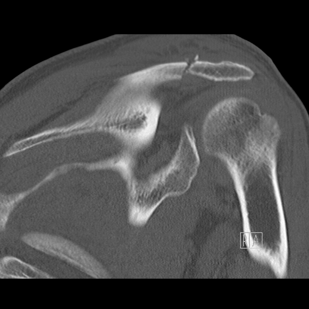
Computed tomography is sometimes necessary when the shoulder radiographs are normal and when there is high clinical suspicion of acromial injury.
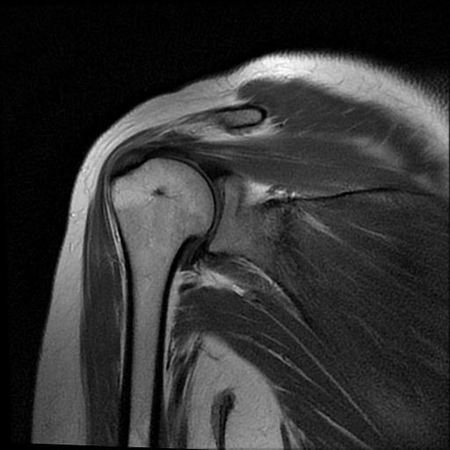
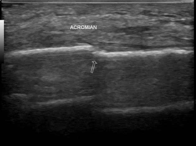
MRI and US exams are also helpful in the assessment of the soft tissues of the shoulder region 4,6.
Classification
Three types of acromial fracture are defined by Kuhn et al 7 which can help to determine whether surgical or non-surgical treatment is appropriate:
-
type I: non- or minimally displaced
IA: avulsion fractures
IB: true fractures
type II: displaced but does not reduce the subacromial space
-
type III: displaced with narrowing of the subacromial space
due to inferior displacement of the acromium; or
superior displacement of an associated glenoid neck fracture
Treatment and prognosis
Type I and II acromial fractures are usually managed with non-surgical treatment while type III fractures usually require surgery to prevent secondary impingement. A variety of surgical techniques can be used and include 7,8:
tension band wiring
reconstruction plate
Kirshner wire
Differential diagnosis
Os acromiale is an unfused acromion accessory ossification center which is relatively common and found in ~8% of the population 9,10. It is bilateral in ~ 60% of individuals 11.

References
- 1. Kenneth A. Egol, Kenneth J. Koval, Joseph David Zuckerman. Handbook of Fractures. (2019) ISBN: 9781605477602
- 2. Charles A. Rockwood, Robert W. Bucholz, Charles M. Court-Brown, James D. Heckman, Paul Tornetta. Rockwood and Green's Fractures in Adults. (2019) ISBN: 9781605476773
- 3. Goodrich JA, Crosland E, Pye J. Acromion fracture associated with posterior shoulder dislocation. (1998) Journal of orthopaedic trauma. 12 (7): 521-3. doi:10.1097/00005131-199809000-00018 - Pubmed
- 4. Getz C, Deutsch A, Williams Junior GR. Scapular and glenoid fractures. In: Jon J. P. Warner, Joseph P. Iannotti, Evan L. Flatow. Complex and Revision Problems in Shoulder Surgery. (2005) ISBN: 9780781746588 - Google Books. pp. 378-80.
- 5. Jamie Weir, Peter H. Abrahams. Imaging Atlas of Human Anatomy. (2019) ISBN: 9780723434573
- 6. Kuhn JE, Blasier RB, Carpenter JE. Fractures of the acromion process: a proposed classification system. (1994) Journal of orthopaedic trauma. 8 (1): 6-13. doi:10.1097/00005131-199402000-00002 - Pubmed
- 7. Phoebe Kaplan. Musculoskeletal MRI. (2001) ISBN: 9780721690278 - Google Books
- 8. Athanasios Papatheodorou, Panagiotis Ellinas, Fotios Takis, Antonios Tsanis, Ioannis Maris, Nikolaos Batakis. US of the Shoulder: Rotator Cuff and Non–Rotator Cuff Disorders1. (2006) RadioGraphics. 26 (1): e23. doi:10.1148/rg.e23 - Pubmed
- 9. Michael B. Zlatkin. MRI of the Shoulder. (2019) ISBN: 9780781715904
Incoming Links
Related articles: Fractures
-
fracture
- terminology
- fracture location
- diaphyseal fracture
- metaphyseal fracture
- physeal fracture
- epiphyseal fracture
- fracture types
- avulsion fracture
- articular surface injuries
- complete fracture
- incomplete fracture
- infraction
- compound fracture
- pathological fracture
- stress fracture
- fracture displacement
- fracture location
- fracture healing
- skull fractures
-
facial fractures
- fractures involving a single facial buttress
- alveolar process fractures
- frontal sinus fracture
- isolated zygomatic arch fractures
- mandibular fracture
- nasal bone fracture
- orbital blow-out fracture
- paranasal sinus fractures
- complex fractures
- dental fractures
- fractures involving a single facial buttress
-
spinal fractures
- classification (AO Spine classification systems)
-
cervical spine fracture classification systems
- AO classification of upper cervical injuries
- AO classification of subaxial injuries
- Anderson and D'Alonzo classification (odontoid fracture)
- Roy-Camille classification (odontoid process fracture)
- Gehweiler classifcation (atlas fractures)
- Levine and Edwards classification (hangman fracture)
- Allen and Ferguson classification (subaxial spine injuries)
- subaxial cervical spine injury classification (SLIC)
- thoracolumbar spinal fracture classification systems
- three column concept of spinal fractures (Denis classification)
- classification of sacral fractures
-
cervical spine fracture classification systems
- spinal fractures by region
- spinal fracture types
- classification (AO Spine classification systems)
- rib fractures
- sternal fractures
-
upper limb fractures
- classification
- Rockwood classification (acromioclavicular joint injury)
- AO classification (clavicle fracture)
- Neer classification (clavicle fracture)
- Neer classification (proximal humeral fracture)
- AO classification (proximal humeral fracture)
- AO/OTA classification of distal humeral fractures
- Milch classification (lateral humeral condyle fracture)
- Weiss classification (lateral humeral condyle fracture)
- Bado classification of Monteggia fracture-dislocations (radius-ulna)
- Mason classification (radial head fracture)
- Frykman classification (distal radial fracture)
- Mayo classification (scaphoid fracture)
- Hintermann classification (gamekeeper's thumb)
- Eaton classification (volar plate avulsion injury)
- Keifhaber-Stern classification (volar plate avulsion injury)
- upper limb fractures by region
- shoulder
- clavicular fracture
-
scapular fracture
- acromion fracture
- coracoid process fracture
- glenoid fracture
- humeral head fracture
- proximal humeral fracture
- humeral neck fracture
- arm
- elbow
- forearm
- wrist
-
carpal bones
- scaphoid fracture
- lunate fracture
- capitate fracture
- triquetral fracture
- pisiform fracture
- hamate fracture
- trapezoid fracture
- trapezium fracture
- hand
- shoulder
- classification
- lower limb fractures
- classification by region
- pelvic fractures
- hip fractures
- Pipkin classification (femoral head fracture)
- Garden classification (hip fracture)
- American Academy of Orthopedic Surgeons classification (periprosthetic hip fracture)
- Cooke and Newman classification (periprosthetic hip fracture)
- Johansson classification (periprosthetic hip fracture)
- Vancouver classification (periprosthetic hip fracture)
- femoral
- knee
- Schatzker classification (tibial plateau fracture)
- AO classification of distal femur fractures
- Meyers and McKeevers classification (anterior cruciate ligament avulsion fracture)
- tibia/fibula
- Watson-Jones classification (tibial tuberosity avulsion fracture)
- ankle
- foot
- Berndt and Harty classification (osteochondral lesions of the talus)
- Sanders CT classification (calcaneal fracture)
- Hawkins classification (talar neck fracture)
- Myerson classification (Lisfranc injury)
- Nunley-Vertullo classification (Lisfranc injury)
- pelvis and lower limb fractures by region
- pelvic fracture
- sacral fracture
- coccygeal fracture
-
hip
- acetabular fracture
- femoral head fracture
-
femoral neck fracture
- subcapital fracture
- transcervical fracture
- basicervical fracture
-
trochanteric fracture
- pertrochanteric fracture
- intertrochanteric fracture
- subtrochanteric fracture
- femur
- mid-shaft fracture
- bisphosphonate-related fracture
- distal femoral fracture
- knee
- avulsion fractures
- Segond fracture
- reverse Segond fracture
- anterior cruciate ligament avulsion fracture
- posterior cruciate ligament avulsion fracture
- arcuate complex avulsion fracture (arcuate sign)
- biceps femoris avulsion fracture
- iliotibial band avulsion fracture
- semimembranosus tendon avulsion fracture
- Stieda fracture (MCL avulsion fracture)
- patellar fracture
- tibial plateau fracture
- avulsion fractures
- leg
- tibial tuberosity avulsion fracture
- tibial shaft fracture
- fibular shaft fracture
- Maisonneuve fracture
- ankle
- foot
- tarsal bones
- metatarsal bones
- phalanges
- classification by region
- terminology