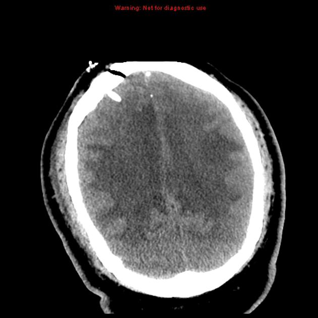
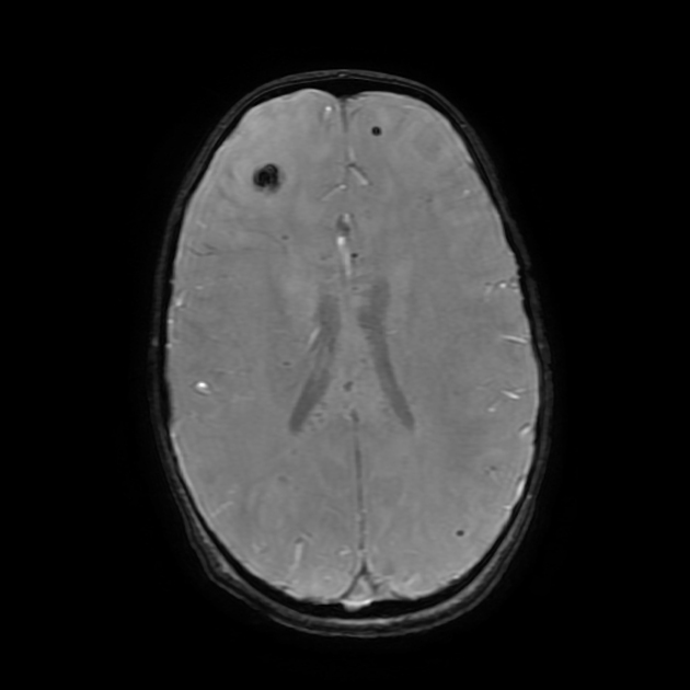
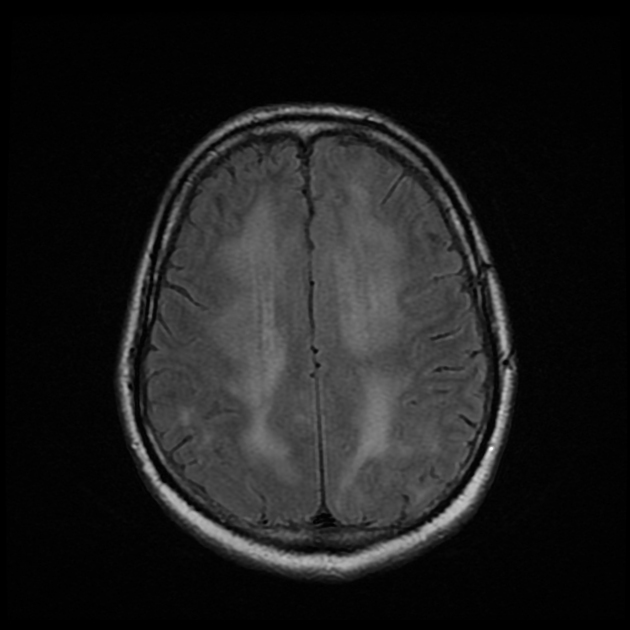
Acute haemorrhagic encephalomyelitis (AHEM), also known as acute haemorrhagic leukoencephalitis (AHLE), Hurst disease or Weston Hurst syndrome, is a very rare form of demyelinating disease. It occurs sporadically and may be considered as the most severe form of acute disseminated encephalomyelitis (ADEM) and is characterised by an acute rapidly progressive fulminant inflammation of the white matter. The cause is unclear but may be post-infectious (either viral or bacterial).
On this page:
Epidemiology
AHEM may more commonly occur in young adults, contrary to ADEM, which is more commonly diagnosed in children 5. It has been reported in older adults 6. There is no gender predilection.
Clinical presentation
Patients present with fever, neck stiffness, fatigue, headache, nausea and vomiting, seizures and coma.
A history of upper respiratory infection prior to presenting neurologic symptoms (interval 2 to 12 days) may be present in ~50% of cases 4.
Pathology
AHEM is related to autoimmune cross reaction to the myelin antigens, and is defined by the presence of necrotising venulitis with perivascular haemorrhages, diffuse CNS ischaemic lesions and myelin destruction 4. Many authors consider it a fulminant variant of ADEM, forming a spectrum of disease 4,5.
Macroscopic appearance
marked cerebral oedema and congestion, often with herniation
punctate haemorrhages within the cerebral white matter, frequently necrotic
commonly presence of necrosis in basal ganglia
typically no involvement of the spinal cord
Microscopic appearance
Imaging appearances are dominated by fibrinoid venous necrosis, leading to acute haemorrhage with fibrin exudates and neutrophilic debris:
classically haemorrhages around the necrotic venules resembling “ring and ball”
white matter ischaemic changes adjacent to necrotic postcapillary venules
haemorrhage more prominent than demyelination
no specific immunohistochemical pattern 4
Radiographic features
MRI
This is the gold-standard and may depict aforementioned morphologic changes such as:
large tumefactive lesions involving the white matter and sparing the cortex
associated punctate haemorrhages and extensive mass effect and surrounding oedema.
possible involvement of ganglia and thalami
Detection of cerebral microhaemorrhages by GRE or the more sensitive susceptibility weighted images (SWI) is an important finding and may allow for differentiation from ADEM.
Treatment and prognosis
Acute haemorrhagic leukoencephalitis is associated with a very poor prognosis with the majority of affected individuals succumbing to the disease.
History and etymology
It was first described by Edward Weston Hurst (1900-1980), a British neurologist, in 1941 2.
Differential diagnosis
General imaging differential considerations include:
acute disseminated encephalomyelitis (ADEM): probably the milder form of the same disease
-
severe demyelination / multiple sclerosis
acute malignant Marburg type multiple sclerosis
-
cortical involvement is more suggestive of HSV encephalitis


 Unable to process the form. Check for errors and try again.