
An adrenal collision tumour or collision tumour of the adrenal gland is an uncommon condition where two histologically distinct tumours abut each other or are in close proximity in the same adrenal gland.
On this page:
Pathology
Collision tumours have been reported in nearly every organ, for example, collision tumour of the ovary. They may be composed of two primary tumours derived from the same organ, or a primary tumour and a metastasis. Given the high prevalence of adrenal adenoma, this is unsurprisingly the most often encountered component in adrenal collision tumours.
In terms of classification, collision tumours need to be distinguished from a composite tumour, where two different histologic types are intermixed.
Clinical significance
The close approximation of two tumours not intermixed at their interface can create both diagnostic and therapeutic difficulties. A mixed benign and malignant collision tumour can render the malignant component unidentifiable on biopsy, with suboptimal or inappropriate treatment being the obvious consequence. It is, therefore, crucial to ensure that the lesion suspicious for malignancy be sampled amply.
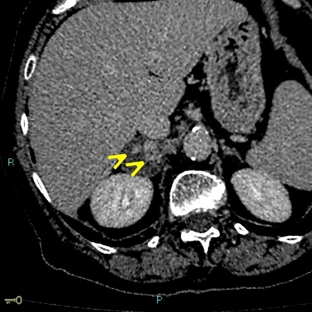
Radiographic assessment
It has been demonstrated that 18F-FDG PET, CT and MRI are all capable of readily distinguishing the two separate components of collision tumours within the adrenal gland. Chemical shift MRI may help in differentiating between benign lipid-poor adenomas and malignant tumours.
In the case of one component being an adrenal adenoma, definitive diagnosis of this component should be straightforward, given the high diagnostic accuracy of the aforementioned modalities for this lesion.
Differential diagnosis
For a heterogeneous appearing adrenal mass consider:
- haemorrhage into a pre-existing non-collision adrenal tumour 3
- adrenal adenoma
- adrenal myelolipoma
- adrenal carcinoma
- adrenal metastases and lymphoma


 Unable to process the form. Check for errors and try again.