
Tuberculous adrenalitis is the result of adrenal Mycobacterium tuberculosis (TB) infection and is part of the spectrum of abdominal TB. Its incidence has decreased in the western world with the declining incidence of tuberculosis.
On this page:
Pathology
As the tuberculous infection causes destruction of the adrenal cortex, primary adrenal insufficiency develops. Pathology usually reveals tuberculous granuloma, caseous necrosis, fibrosis, and calcification.
As adrenal involvement progresses, physiological tests for adrenal insufficiency including plasma/urinary cortical measurement and ACTH challenges can raise suspicious for adrenal TB.
Clinical presentation
Symptoms of adrenal insufficiency may occur, such as fatigue and abdominal pain. When >90% of the cortex has been destroyed, patients may present with Addisonian crisis, which can be life-threatening.
Radiographic features
CT forms the mainstay of evaluation due to its high spatial resolution and availability, but MRI also has a known role in assessing adrenal lesions, particularly in young patients where radiation dose is a concern.
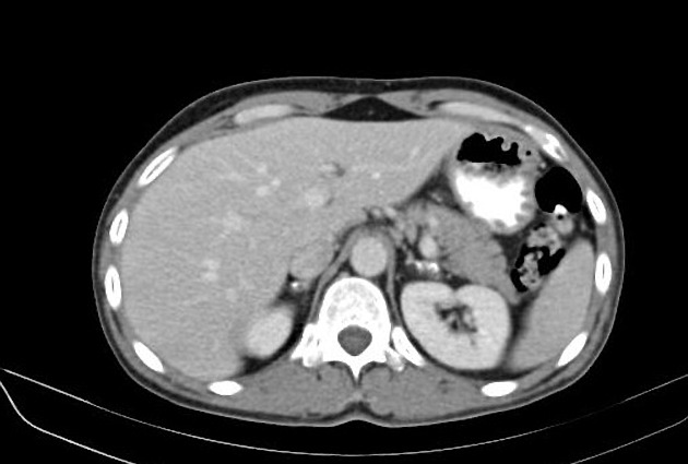
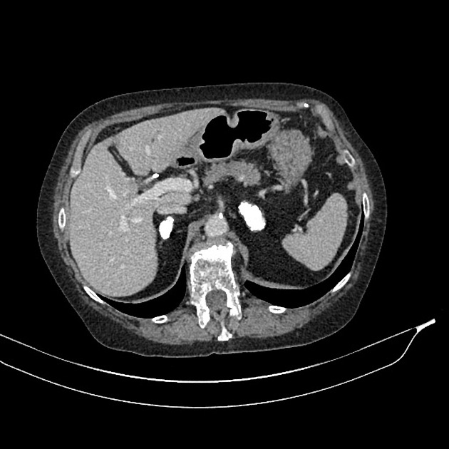
CT
Unenhanced and portal venous phase IV contrast scans are usually performed. A narrow field of view further helps in optimising spatial resolution in detecting discrete lesions.
-
gland contour:
-
in the early stage there can be mass-like adrenal enlargement
smooth adrenal contour is preserved
-
later on adrenal fibrosis and atrophy occurs
small adrenals with irregular margins
-
-
calcification:
this is a late feature, often occurring post-treatment
it can be punctate, localised, or diffuse
-
gland density:
-
central low density can be seen in early disease
due to caseous necrosis
with anti-TB treatment the adrenals show homogenous density
-
-
enhancement:
can see areas of relative central hypoenhancement
MRI
Imaging features are analogous to CT except for MR limitations in assessing calcified tissue.


 Unable to process the form. Check for errors and try again.