
Aliasing on MRI
Citation, DOI, disclosures and article data
At the time the article was created J Yeung had no recorded disclosures.
View J Yeung's current disclosuresAt the time the article was last revised Amanda Er had no financial relationships to ineligible companies to disclose.
View Amanda Er's current disclosures- MRI aliasing
- Wrap around artifact
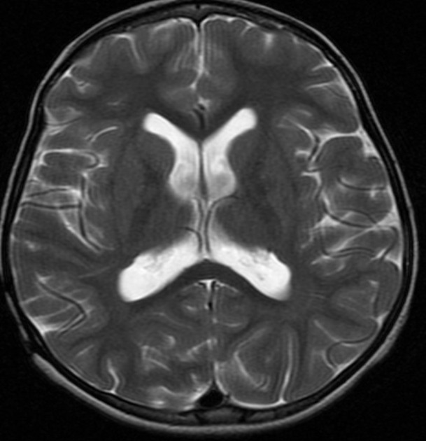
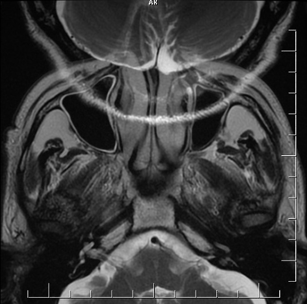
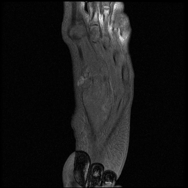
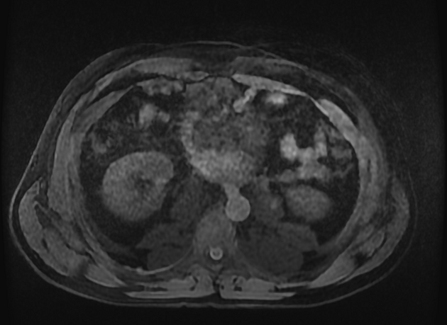
Aliasing on MRI, also known as wrap-around, is a frequently encountered MRI artifact that occurs when the field of view (FOV) is smaller than the body part being imaged. The part of the body that lies beyond the edge of the FOV is projected onto the other side of the image 5.
This can be corrected, if necessary, by oversampling the data. In the frequency direction, this is accomplished by sampling the signal twice as fast. In the phase direction, the number of phase-encoding steps must be increased with a longer study. However, if the FOV and matrix size (phase-encoding steps) are increased and simultaneously the number of excitations (or number of signal averages) reduced to half, the imaging time can be kept constant with correction of aliasing 5.
On this page:
More detail
The basis of aliasing lies in "analogue-to-digital conversion", wherein the continuous MR signal picked by the receiver coil is converted into its digital counterpart for presentation as a grey-scale image. This ubiquitously involves the sampling of the continuous signal at pre-defined intervals. For greater fidelity in signal conversion, the sampling rate should be at least twice the highest frequency within the signal (Nyquist rate). At lower sampling rates, high-frequency signals become indistinguishable from lower frequency signals, i.e. they become aliases 5.
On MRI, spatial localisation within a single image depends on the frequency signature of the MR signal originating from that portion. Within a given bandwidth, higher frequency signals come from the periphery of the image and are aliased over the lower frequency (relatively) central portion of the image. Aliasing on MRI can occur in both phase and frequency axis.
Remedy
Aliasing on MRI can be compensated for by 5:
enlarging the field of view (FOV)
using pre-saturation bands on areas outside the FOV
anti-aliasing software
switching the phase and frequency directions
use a surface coil to reduce the signal outside of the area of interest
See also
References
- 1. Morelli J, Runge V, Ai F et al. An Image-Based Approach to Understanding the Physics of MR Artifacts. Radiographics. 2011;31(3):849-66. doi:10.1148/rg.313105115 - Pubmed
- 2. Pusey E, Lufkin R, Brown R et al. Magnetic Resonance Imaging Artifacts: Mechanism and Clinical Significance. Radiographics. 1986;6(5):891-911. doi:10.1148/radiographics.6.5.3685515 - Pubmed
- 3. Catherine Westbrook, Carolyn Kaut Roth, John Talbot. MRI in Practice. (2011) ISBN: 9781444337433 - Google Books
- 4. Penelope Allisy-Roberts, Jerry R. Williams. Farr's Physics for Medical Imaging. (2007) ISBN: 9780702028441 - Google Books
- 5. Kaur P, Senthil Kumaran S, Tripathi R, Khushu S, Kaushik S. Protocol Error Artifacts in MRI: Sources and Remedies Revisited. Radiography. 2007;13(4):291-306. doi:10.1016/j.radi.2006.03.011
Incoming Links
Related articles: Imaging technology
- imaging technology
- imaging physics
- imaging in practice
-
x-rays
- x-ray physics
- x-ray in practice
- x-ray production
- x-ray tube
- filters
- automatic exposure control (AEC)
- beam collimators
- grids
- air gap technique
- cassette
- intensifying screen
- x-ray film
- image intensifier
- digital radiography
- digital image
- mammography
- x-ray artifacts
- radiation units
- radiation safety
- radiation detectors
- fluoroscopy
-
computed tomography (CT)
- CT physics
- CT in practice
- CT technology
- CT image reconstruction
- CT image quality
- CT dose
-
CT contrast media
-
iodinated contrast media
- agents
- water soluble
- water insoluble
- vicarious contrast material excretion
- iodinated contrast media adverse reactions
- agents
- non-iodinated contrast media
-
iodinated contrast media
-
CT artifacts
- patient-based artifacts
- physics-based artifacts
- hardware-based artifacts
- ring artifact
- tube arcing
- out of field artifact
- air bubble artifact
- helical and multichannel artifacts
- CT safety
- history of CT
-
MRI
- MRI physics
- MRI in practice
- MRI hardware
- signal processing
-
MRI pulse sequences (basics | abbreviations | parameters)
- T1 weighted image
- T2 weighted image
- proton density weighted image
- chemical exchange saturation transfer
- CSF flow studies
- diffusion weighted imaging (DWI)
- echo-planar pulse sequences
- fat-suppressed imaging sequences
- gradient echo sequences
- inversion recovery sequences
- metal artifact reduction sequence (MARS)
-
perfusion-weighted imaging
- techniques
- derived values
- saturation recovery sequences
- spin echo sequences
- spiral pulse sequences
- susceptibility-weighted imaging (SWI)
- T1 rho
- MR angiography (and venography)
-
MR spectroscopy (MRS)
- 2-hydroxyglutarate peak: resonates at 2.25 ppm
- alanine peak: resonates at 1.48 ppm
- choline peak: resonates at 3.2 ppm
- citrate peak: resonates at 2.6 ppm
- creatine peak: resonates at 3.0 ppm
- functional MRI (fMRI)
- gamma-aminobutyric acid (GABA) peak: resonates at 2.2-2.4 ppm
- glutamine-glutamate peak: resonates at 2.2-2.4 ppm
- Hunter's angle
- lactate peak: resonates at 1.3 ppm
- lipids peak: resonates at 1.3 ppm
- myoinositol peak: resonates at 3.5 ppm
- MR fingerprinting
- N-acetylaspartate (NAA) peak: resonates at 2.0 ppm
- propylene glycol peak: resonates at 1.13 ppm
-
MRI artifacts
- MRI hardware and room shielding
- MRI software
- patient and physiologic motion
- tissue heterogeneity and foreign bodies
- Fourier transform and Nyquist sampling theorem
- MRI contrast agents
- MRI safety
-
ultrasound
- ultrasound physics
-
transducers
- linear array
- convex array
- phased array
- frame averaging (frame persistence)
- ultrasound image resolution
- imaging modes and display
- pulse-echo imaging
- real-time imaging
-
Doppler imaging
- Doppler effect
- colour Doppler
- power Doppler
- B flow
- colour box
- Doppler angle
- pulse repetition frequency and scale
- wall filter
- colour write priority
- packet size (dwell time)
- peak systolic velocity
- end-diastolic velocity
- resistive index
- pulsatility index
- Reynolds number
- panoramic imaging
- compound imaging
- harmonic imaging
- elastography
- scanning modes
- 2D ultrasound
- 3D ultrasound
- 4D ultrasound
- M-mode
-
ultrasound artifacts
- acoustic shadowing
- acoustic enhancement
- beam width artifact
- reverberation artifact
- ring down artifact
- mirror image artifact
- side lobe artifact
- speckle artifact
- speed displacement artifact
- refraction artifact
- multipath artifact
- anisotropy
- electrical interference artifact
- hardware-related artifacts
- Doppler artifacts
- aliasing
- tissue vibration
- spectral broadening
- blooming
- motion (flash) artifact
- twinkling artifact
- acoustic streaming
- biological effects of ultrasound
- history of ultrasound
-
nuclear medicine
- nuclear medicine physics
- detectors
- tissue to background ratio
-
radiopharmaceuticals
- fundamentals of radiopharmaceuticals
- radiopharmaceutical labelling
- radiopharmaceutical production
- nuclear reactor produced radionuclides
- cyclotron produced radionuclides
- radiation detection
- dosimetry
- specific agents
- carbon-11
- chromium-51
- fluorine agents
- gallium agents
- Ga-67 citrate
- Ga-68
- iodine agents
-
I-123
- I-123 iodide
- I-123 ioflupane (DaTSCAN)
- I-123 ortho-iodohippurate
- I-131
-
MIBG scans
- I-123 MIBG
- I-131 MIBG
-
I-123
- indium agents
- In-111 Octreoscan
- In-111 OncoScint
- In-111 Prostascint
- In-111 oxine labelled WBC
- krypton-81m
- nitrogen-13
- oxygen-15
- phosphorus-32
- selenium-75
-
technetium agents
- Tc-99m DMSA
- Tc-99m DTPA
- Tc-99m DTPA aerosol
- Tc-99m HMPAO
- Tc-99m HMPAO labelled WBC
- Tc-99m MAA
- Tc-99m MAG3
- Tc-99m MDP
- Tc-99m mercaptoacetyltriglycine
- Tc-99m pertechnetate
- Tc-99m labelled RBC
- Tc-99m sestamibi
- Tc-99m sulfur colloid
- Tc-99m sulfur colloid (oral)
- thallium-201 chloride
- xenon agents
- in vivo therapeutic agents
- pharmaceuticals used in nuclear medicine
-
emerging methods in medical imaging
- radiography
- phase-contrast imaging
- CT
- deep-learning reconstruction
- photon counting CT
- virtual non-contrast imaging
- ultrasound
- magnetomotive ultrasound (MMUS)
- superb microvascular imaging
- ultrafast Doppler imaging
- ultrasound localisation microscopy
- MRI
- nuclear medicine
- total body PET system
- immuno-PET
- miscellaneous
- radiography


 Unable to process the form. Check for errors and try again.