
Anderson and Montesano classification of occipital condyle fractures
Citation, DOI, disclosures and article data
Citation:
Hacking C, Deng F, Anderson and Montesano classification of occipital condyle fractures. Reference article, Radiopaedia.org (Accessed on 21 Mar 2025) https://doi.org/10.53347/rID-87202
rID:
87202
Article created:
Disclosures:
At the time the article was created Craig Hacking had no recorded disclosures.
View Craig Hacking's current disclosures
Last revised:
Disclosures:
At the time the article was last revised Francis Deng had no recorded disclosures.
View Francis Deng's current disclosures
Revisions:
5 times, by
2 contributors -
see full revision history and disclosures
Systems:
Sections:
Synonyms:
- Anderson-Montesano classification of occipital condyle fractures
The Anderson and Montesano classification is a widely used system for describing occipital condyle fractures. It divides injuries into three types based on morphology and mechanism of injury 1-5.
On this page:
Classification
-
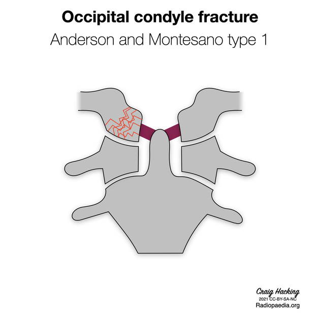
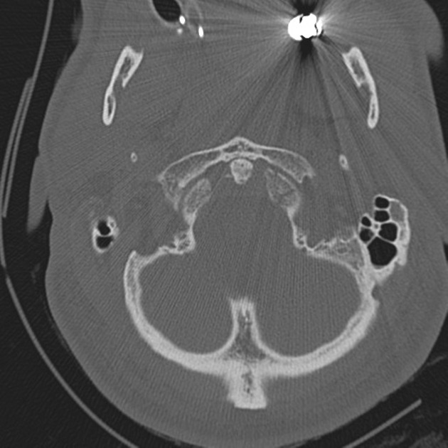
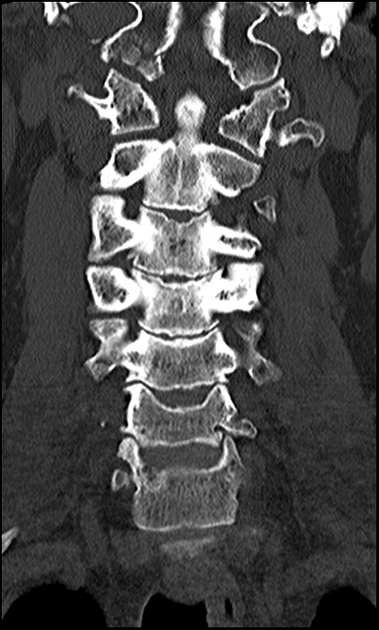
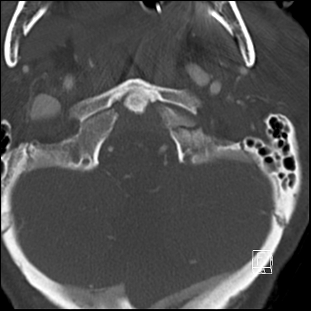
type I: impacted type occipital condyle fracture
- morphology: comminution of the condyle with minimal or no displacement of fragments into the foramen magnum
- mechanism: axial loading of the skull onto the atlas
- stability: stable because the tectorial membrane and contralateral alar ligament are intact (the ipsilateral alar ligament may be functionally inadequate)
-
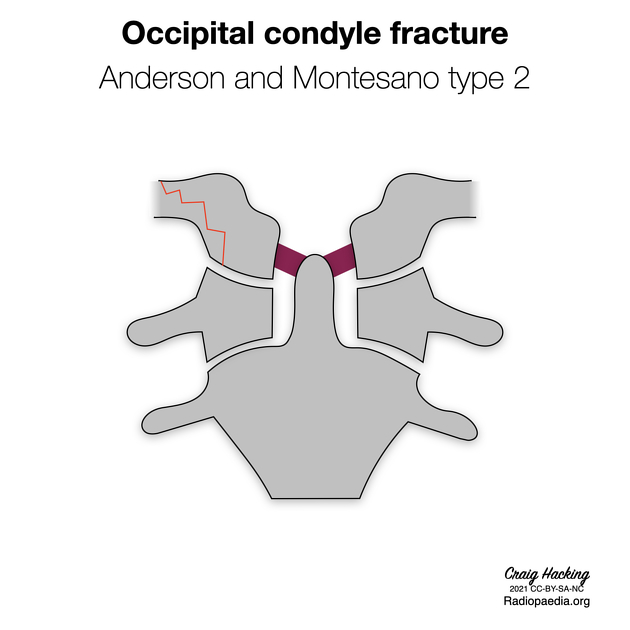
type II: basilar skull type occipital condyle fracture
- morphology: condyle fracture that extends outside of the condyle to elsewhere in the posterior base of skull
- mechanism: direct blow to the lower skull
- stability: stable because the tectorial membrane and alar ligaments are intact
-
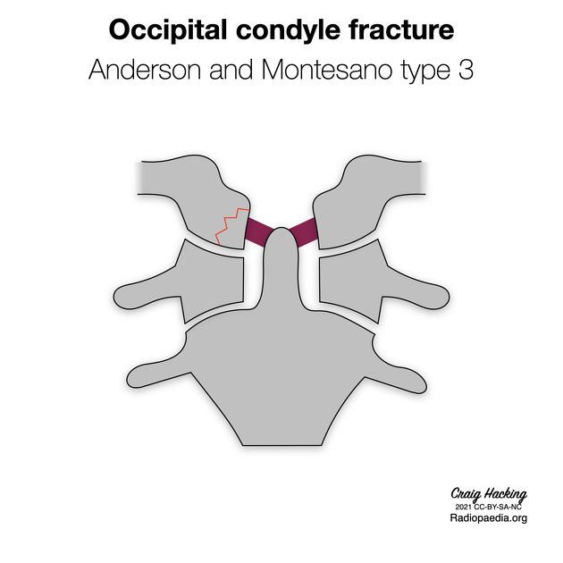
type III: avulsion type occipital condyle fracture
- morphology: small inferomedial occipital condyle fracture fragment displaced toward the odontoid tip
- mechanism: rotation and/or contralateral bending
- stability: potentially unstable due to loading of the contralateral alar ligament and tectorial membrane
Epidemiology
A range of incidences of the Anderson and Montesano types amongst all occipital condyle fractures has been reported 2,4,6:
- type I: 3-13%
- type II: 22-54%
- type III: 33-75%
History and etymology
The classification was described in 1988 by American orthopaedic surgeons Paul A. Anderson and Pasquale X. Montesano on the basis of 6 patients diagnosed by conventional anterior posterior polytomography or CT with coronal reconstructions 1.
See also
References
- 1. Anderson P & Montesano P. Morphology and Treatment of Occipital Condyle Fractures. Spine (Phila Pa 1976). 1988;13(7):731-6. doi:10.1097/00007632-198807000-00004 - Pubmed
- 2. Noble E & Smoker W. The Forgotten Condyle: The Appearance, Morphology, and Classification of Occipital Condyle Fractures. AJNR Am J Neuroradiol. 1996;17(3):507-13. PMC8337992 - Pubmed
- 3. Leone A, Cerase A, Colosimo C, Lauro L, Puca A, Marano P. Occipital Condylar Fractures: A Review. Radiology. 2000;216(3):635-44. doi:10.1148/radiology.216.3.r00se23635 - Pubmed
- 4. Hanson J, Deliganis A, Baxter A et al. Radiologic and Clinical Spectrum of Occipital Condyle Fractures. AJR Am J Roentgenol. 2002;178(5):1261-8. doi:10.2214/ajr.178.5.1781261 - Pubmed
- 5. Riascos R, Bonfante E, Cotes C, Guirguis M, Hakimelahi R, West C. Imaging of Atlanto-Occipital and Atlantoaxial Traumatic Injuries: What the Radiologist Needs to Know. Radiographics. 2015;35(7):2121-34. doi:10.1148/rg.2015150035 - Pubmed
- 6. Aulino J, Tutt L, Kaye J, Smith P, Morris J. Occipital Condyle Fractures: Clinical Presentation and Imaging Findings in 76 Patients. Emerg Radiol. 2005;11(6):342-7. doi:10.1007/s10140-005-0425-0 - Pubmed
Incoming Links
Articles:
Cases:
- Occipital condyle fracture (type 2) with extension into clivus
- Bilateral occipital condyle fracture (type 2)
- Occipital condyle fracture (type 1) and atlas transverse process fracture (type 5)
- Occipital condyle fracture (type 3)
- Bilateral occipital condyle fractures (type 3)
- Occipital condyle fracture (type 1)
- Anderson and Montesano classification of occipital condyle fractures (diagrams)
Related articles: Fractures
-
fracture
- terminology
- fracture location
- diaphyseal fracture
- metaphyseal fracture
- physeal fracture
- epiphyseal fracture
- fracture types
- avulsion fracture
- articular surface injuries
- complete fracture
- incomplete fracture
- infraction
- compound fracture
- pathological fracture
- stress fracture
- fracture displacement
- fracture location
- fracture healing
- skull fractures
-
facial fractures
- fractures involving a single facial buttress
- alveolar process fractures
- frontal sinus fracture
- isolated zygomatic arch fractures
- mandibular fracture
- nasal bone fracture
- orbital blow-out fracture
- paranasal sinus fractures
- complex fractures
- dental fractures
- fractures involving a single facial buttress
-
spinal fractures
- classification (AO Spine classification systems)
-
cervical spine fracture classification systems
- AO classification of upper cervical injuries
- AO classification of subaxial injuries
- Anderson and D'Alonzo classification (odontoid fracture)
- Roy-Camille classification (odontoid process fracture)
- Gehweiler classifcation (atlas fractures)
- Levine and Edwards classification (hangman fracture)
- Allen and Ferguson classification (subaxial spine injuries)
- subaxial cervical spine injury classification (SLIC)
- thoracolumbar spinal fracture classification systems
- three column concept of spinal fractures (Denis classification)
- classification of sacral fractures
-
cervical spine fracture classification systems
- spinal fractures by region
- spinal fracture types
- classification (AO Spine classification systems)
- rib fractures
- sternal fractures
-
upper limb fractures
- classification
- Rockwood classification (acromioclavicular joint injury)
- AO classification (clavicle fracture)
- Neer classification (clavicle fracture)
- Neer classification (proximal humeral fracture)
- AO classification (proximal humeral fracture)
- AO/OTA classification of distal humeral fractures
- Milch classification (lateral humeral condyle fracture)
- Weiss classification (lateral humeral condyle fracture)
- Bado classification of Monteggia fracture-dislocations (radius-ulna)
- Mason classification (radial head fracture)
- Frykman classification (distal radial fracture)
- Mayo classification (scaphoid fracture)
- Hintermann classification (gamekeeper's thumb)
- Eaton classification (volar plate avulsion injury)
- Keifhaber-Stern classification (volar plate avulsion injury)
- upper limb fractures by region
- shoulder
- clavicular fracture
-
scapular fracture
- acromion fracture
- coracoid process fracture
- glenoid fracture
- humeral head fracture
- proximal humeral fracture
- humeral neck fracture
- arm
- elbow
- forearm
- wrist
-
carpal bones
- scaphoid fracture
- lunate fracture
- capitate fracture
- triquetral fracture
- pisiform fracture
- hamate fracture
- trapezoid fracture
- trapezium fracture
- hand
- shoulder
- classification
- lower limb fractures
- classification by region
- pelvic fractures
- hip fractures
- Pipkin classification (femoral head fracture)
- Garden classification (hip fracture)
- American Academy of Orthopaedic Surgeons classification (periprosthetic hip fracture)
- Cooke and Newman classification (periprosthetic hip fracture)
- Johansson classification (periprosthetic hip fracture)
- Vancouver classification (periprosthetic hip fracture)
- femoral
- knee
- Schatzker classification (tibial plateau fracture)
- AO classification of distal femur fractures
- Meyers and McKeevers classification (anterior cruciate ligament avulsion fracture)
- tibia/fibula
- Watson-Jones classification (tibial tuberosity avulsion fracture)
- ankle
- foot
- Berndt and Harty classification (osteochondral lesions of the talus)
- Sanders CT classification (calcaneal fracture)
- Hawkins classification (talar neck fracture)
- Myerson classification (Lisfranc injury)
- Nunley-Vertullo classification (Lisfranc injury)
- pelvis and lower limb fractures by region
- pelvic fracture
- sacral fracture
- coccygeal fracture
-
hip
- acetabular fracture
- femoral head fracture
-
femoral neck fracture
- subcapital fracture
- transcervical fracture
- basicervical fracture
-
trochanteric fracture
- pertrochanteric fracture
- intertrochanteric fracture
- subtrochanteric fracture
- femur
- mid-shaft fracture
- bisphosphonate-related fracture
- distal femoral fracture
- knee
- avulsion fractures
- Segond fracture
- reverse Segond fracture
- anterior cruciate ligament avulsion fracture
- posterior cruciate ligament avulsion fracture
- arcuate complex avulsion fracture (arcuate sign)
- biceps femoris avulsion fracture
- iliotibial band avulsion fracture
- semimembranosus tendon avulsion fracture
- Stieda fracture (MCL avulsion fracture)
- patellar fracture
- tibial plateau fracture
- avulsion fractures
- leg
- tibial tuberosity avulsion fracture
- tibial shaft fracture
- fibular shaft fracture
- Maisonneuve fracture
- ankle
- foot
- tarsal bones
- metatarsal bones
- phalanges
- classification by region
- terminology
Related articles: Spinal trauma
-
spinal fractures
- morphology
- fractures by location
- cervical spine fracture
- thoracolumbar spine fracture
- sacral fracture
- classifications
- AO spine classification systems
- three column concept of spinal fractures (Denis classification)
-
cervical spine fracture classification systems
- upper cervical spine
- AO Spine classification of upper cervical injuries
- occipital condyle and occipital cervical junction
- atlas (C1) and C1-2 joint
- axis (C2) and C2-3 joint
- Roy-Camille classification (dens)
- Anderson and D'Alonzo classification (dens)
- Levine and Edwards classification (pars interarticularis)
- subaxial cervical spine
- upper cervical spine
- thoracolumbar spinal fracture classification systems
- classifications of sacral fractures
- facet dislocation
- listhesis


 Unable to process the form. Check for errors and try again.