
Anterior longitudinal ligament
Citation, DOI, disclosures and article data
Citation:
Wong A, Hacking C, Haouimi A, et al. Anterior longitudinal ligament. Reference article, Radiopaedia.org (Accessed on 27 Mar 2025) https://doi.org/10.53347/rID-36568
Permalink:
rID:
36568
Article created:
30 Apr 2015,
Aaron Wong
Disclosures:
At the time the article was created Aaron Wong had no recorded disclosures.
View Aaron Wong's current disclosures
Last revised:
Disclosures:
At the time the article was last revised Craig Hacking had no recorded disclosures.
View Craig Hacking's current disclosures
Revisions:
16 times, by
12 contributors -
see full revision history and disclosures
Systems:
Sections:
Synonyms:
- Anterior longitudinal ligament (ALL)
- Anterior longitudinal ligaments
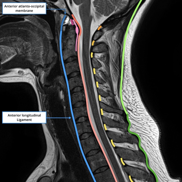
The anterior longitudinal ligament (ALL) runs along the anterior surface of the vertebral bodies (firmly united to the periosteum) and intervertebral discs (attaching to the anterior annulus). It ascends from the anterosuperior portion of the sacrum superiorly to become the anterior atlantooccipital membrane at the level of the anterior arch of C1 (atlas) 1-6.
It can be divided into three distinct layers 2,3.
- superficial: traverses 3-4 vertebrae
- intermediate: covers 2-3 vertebrae
-
deep
- between individual vertebrae, it either blends into the periosteum or inserts anteriorly
- the first ALL layer is typically affected in DISH 4
References
- 1. McMinn. Lasts Anatomy Regional and Applied. Churchill Livingstone (2003) ISBN:B0084AQDG8. Read it at Google Books - Find it at Amazon
- 2. PS Ramani. WFNS Spine Committee Textbook on Thoracic Spine. ISBN: 9789352500079
- 3. Nikolai Bogduk. Clinical and Radiological Anatomy of the Lumbar Spine. ISBN: 9780702051661
- 4. Albert L. Baert. Encyclopedia of Imaging. ISBN: 9783540352785
- 5. Offiah CE, Day E. The craniocervical junction: embryology, anatomy, biomechanics and imaging in blunt trauma. Insights into imaging. 8 (1): 29-47. doi:10.1007/s13244-016-0530-5 - Pubmed
- 6. Walter Carl Hartwig. Fundamental Anatomy. (2018) ISBN: 9780781768887
Incoming Links
Articles:
- Coccyx
- Flowing ossifications
- Intervertebral joint
- Diaphragm
- AO Spine classification of subaxial injuries
- Cervical spine ligaments
- Sacrum
- Medical abbreviations and acronyms (A)
- Azygos vein
- Superior thoracic aperture
- Axis (C2)
- Atlas (C1)
- Extension teardrop fracture
- Infantile cervical ligament oedema
- Atlanto-occipital articulation
- Atlanto-axial articulation
- Diffuse idiopathic skeletal hyperostosis
- Symphysis
- Inferior vena cava
- Lumbar spine
Cases:
- Spinal cord transection
- Ligaments of the lumbar spine (Gray's illustration)
- Extension teardrop fracture
- Diffuse idiopathic skeletal hyperotosis
- Ligament injury and cord oedema in cervical spine trauma
- Cervical facet fracture and anterior discoligamentous injury
- Diffuse idiopathic skeletal hyperostosis (DISH)
Related articles: Anatomy: Spine
-
osteology
- vertebrae
- spinal canal
- cervical spine
- thoracic spine
- lumbar spine
- sacrum
- coccyx
-
anatomical variants
- vertebral body
- neural arch
- transitional vertebrae
- ossicles
- ossification centres
- intervertebral disc
- articulations
- ligaments
- musculature of the vertebral column
- muscles of the neck
- muscles of the back
-
suboccipital muscle group
- rectus capitis posterior major muscle
- rectus capitis posterior minor muscle
- obliquus capitis superior muscle
- obliquus capitis inferior muscle
- splenius capitis muscle
- splenius cervicis muscle
- erector spinae group
- transversospinalis group
- quadratus lumborum muscle
-
suboccipital muscle group
- spinal meninges and spaces
-
spinal cord
- gross anatomy
-
white matter tracts (white matter)
- corticospinal tract
- anterolateral columns
- lateral columns
-
dorsal columns
- fasiculus gracilis (column of Goll)
- fasiculus cuneatus (column of Burdach)
- grey matter
- nerve root
- central canal
- functional anatomy
- spinal cord blood supply
- sympathetic chain


 Unable to process the form. Check for errors and try again.