
Benedikt syndrome
Citation, DOI, disclosures and article data
At the time the article was created Bruno Di Muzio had no recorded disclosures.
View Bruno Di Muzio's current disclosuresAt the time the article was last revised Mostafa Elfeky had no financial relationships to ineligible companies to disclose.
View Mostafa Elfeky's current disclosures- Paramedian midbrain syndrome
- Benedikt's syndrome
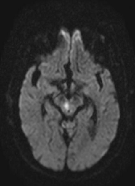
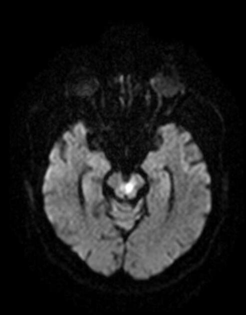
Benedikt syndrome, or paramedian midbrain syndrome, is a midbrain stroke syndrome that involves the fascicles of the oculomotor nerve and the red nucleus.
On this page:
Clinical presentation
- ipsilateral CN III palsy 1-4
- crossed hemiataxia 1-4
- crossed choreoathetosis 1-4
Pathology
It is usually caused by an ischaemic stroke, typically involving branches of the posterior cerebral artery 1-4.
Radiographic features
Using imaging alone, it is difficult to distinguish Benedikt syndrome from Weber syndrome, unless clear involvement of the red nucleus can be identified, which is seen in the former 1-4.
History and etymology
The syndrome was first described by Moritz Benedikt (1835-1920), a Hungarian-Austrian neurologist, in 1889 5.
References
- 1. Jacobs DA, Galetta SL. Neuro-ophthalmology for neuroradiologists. AJNR Am J Neuroradiol. 2007;28 (1): 3-8. Pubmed citation
- 2. Cormier PJ, Long ER, Russell EJ. MR imaging of posterior fossa infarctions: vascular territories and clinical correlates. Radiographics. 1992;12 (6): 1079-96. doi:10.1148/radiographics.12.6.1439013 - Pubmed citation
- 3. Liu GT, Crenner CW, Logigian EL, Charness ME, Samuels MA. Midbrain syndromes of Benedikt, Claude, and Nothnagel: setting the record straight. Neurology. 42 (9): 1820-2. Pubmed
- 4. Allan Ropper, Joshua Klein, Martin Samuels. Adams and Victor's Principles of Neurology 10th Edition. (2014) ISBN: 9780071794794 - Google Books
- 5. Benedikt M. Tremblement avec paralysie croisee du moteur oculaire commun. Bull Med Paris 1889;3:547-548.
Incoming Links
Related articles: Stroke and intracranial haemorrhage
-
stroke and intracranial haemorrhage
- general articles
-
ischaemic stroke
- general discussions
- scoring and classification systems
- Alberta stroke program early CT score (ASPECTS)
- ASCOD classification
- Canadian Neurological Scale
- Heidelberg bleeding classification
- NIH Stroke Scale
- Mathew stroke scale
- modified Rankin scale
- Orgogozo Stroke Scale
- Scandinavian Stroke Scale
- thrombolysis in cerebral infarction (TICI) scale
- TOAST classification
- collateral vessel scores
- signs
- by region
- hemispheric infarcts
- frontal lobe infarct
- parietal lobe infarct
- temporal lobe infarct
- occipital lobe infarct
- alexia without agraphia syndrome: PCA
- cortical blindness syndrome (Anton syndrome): top of basilar or bilateral PCA
- Balint syndrome: bilateral PCA
- lacunar infarct
-
thalamic infarct
- artery of Percheron infarct
- Déjerine-Roussy syndrome (thalamic pain syndrome): thalamoperforators of PCA
- top of the basilar syndrome
- striatocapsular infarct
- choroid plexus infarct
- cerebellar infarct
-
brainstem infarct
- midbrain infarct
- Benedikt syndrome: PCA
- Claude syndrome: PCA
- Nothnagel syndrome: PCA
- Weber syndrome: PCA
- Wernekink commissure syndrome
- pontine infarct
- Brissaud-Sicard syndrome
- facial colliculus syndrome
- Gasperini syndrome: basilar artery or AICA
- inferior medial pontine syndrome (Foville syndrome): basilar artery
- lateral pontine syndrome (Marie-Foix syndrome): basilar artery or AICA
- locked-in syndrome: basilar artery
- Millard-Gubler syndrome: basilar artery
- Raymond syndrome: basilar artery
- medullary infarct
- Babinski-Nageotte syndrome
- Cestan-Chenais syndrome
- hemimedullary syndrome (Reinhold syndrome)
- lateral medullary stroke syndrome (Wallenberg syndrome)
- medial medullary syndrome (Déjerine syndrome)
- Opalski syndrome
- midbrain infarct
- acute spinal cord ischaemia syndrome
- hemispheric infarcts
- by vascular territory
- by vessel size
- treatment options
- complications
-
intracranial haemorrhage
-
intra-axial haemorrhage
- signs and formulas
- ABC/2 (volume estimation)
- black hole sign
- blend sign
- cashew nut sign
- CTA spot sign
- island sign
- satellite sign
- swirl sign
- zebra sign
- by type
- by location
- signs and formulas
- extra-axial haemorrhage
- extradural haemorrhage (EDH)
- intralaminar dural haemorrhage
- subdural haemorrhage (SDH)
-
subarachnoid haemorrhage (SAH)
- types
- complications
- grading systems
- subpial haemorrhage
-
intra-axial haemorrhage


 Unable to process the form. Check for errors and try again.