Citation, DOI, disclosures and article data
Citation:
Yap K, Sharma R, Spires R, et al. Biffl scale for blunt cerebrovascular injury. Reference article, Radiopaedia.org (Accessed on 30 Mar 2025) https://doi.org/10.53347/rID-13710
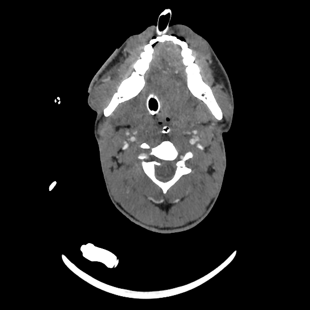
The Biffl scale or grade illustrates the spectrum of blunt cerebrovascular injury (BCVI) seen on angiography (both CTA and DSA). Some authors refer to the grading scale as the Denver scale, which is not to be confused with the Denver criteria, a series of clinical indications to screen for blunt cerebrovascular injury in trauma patients.
Classification
grade I: minimal luminal irregularity or intramural haematoma/dissection with <25% luminal narrowing
grade II: intramural haematoma/dissection with ≥25% luminal narrowing, intraluminal thrombus, or raised intimal flap
grade III: pseudoaneurysm
grade IV: occlusion
grade V: transection with free extravasation
Arteriovenous fistula is often included with transections (grade V) 4,5. However, Biffl et al. initially described fistulae as grade I if small and grade III if large 1, and later revised this description to grade II if small and grade V if hemodynamically significant 6. Other authors have proposed classifying these lesions as grade III 7.
Treatment and prognosis
This grading system has prognostic and therapeutic implications.
The risk of stroke increases with increasing grade of carotid artery injury 1:
grade I: 8%
grade II: 14%
grade III: 26%
grade IV: 50%
grade V: 100%
The risk of stroke does not correlate with increasing grade of vertebral artery injury 3:
-
1. Biffl WL, Moore EE, Offner PJ et-al. Blunt carotid arterial injuries: implications of a new grading scale. J Trauma. 1999;47 (5): 845-53. J Trauma (link) - Pubmed citation
-
2. Cothren CC, Moore EE. Blunt cerebrovascular injuries. Clinics (Sao Paulo). 2006;60 (6): 489-96. Pubmed citation
-
3. Biffl WL, Moore EE, Elliott JP, Ray C, Offner PJ, Franciose RJ, Brega KE, Burch JM. The devastating potential of blunt vertebral arterial injuries. (2000) Annals of surgery. 231 (5): 672-81. doi:10.1097/00000658-200005000-00007 - Pubmed
-
4. Rutman AM, Vranic JE, Mossa-Basha M. Imaging and Management of Blunt Cerebrovascular Injury. (2018) Radiographics : a review publication of the Radiological Society of North America, Inc. 38 (2): 542-563. doi:10.1148/rg.2018170140 - Pubmed
-
5. Nagpal P, Policeni BA, Bathla G, Khandelwal A, Derdeyn C, Skeete D. Blunt Cerebrovascular Injuries: Advances in Screening, Imaging, and Management Trends. (2017) AJNR. American journal of neuroradiology. doi:10.3174/ajnr.A5412 - Pubmed
-
6. Biffl WL, Ray CE, Moore EE, Franciose RJ, Aly S, Heyrosa MG, Johnson JL, Burch JM. Treatment-related outcomes from blunt cerebrovascular injuries: importance of routine follow-up arteriography. (2002) Annals of surgery. 235 (5): 699-706; discussion 706-7. doi:10.1097/00000658-200205000-00012 - Pubmed
-
7. Ares WJ, Jankowitz BT, Tonetti DA, Gross BA, Grandhi R. A comparison of digital subtraction angiography and computed tomography angiography for the diagnosis of penetrating cerebrovascular injury. (2019) Neurosurgical focus. 47 (5): E16. doi:10.3171/2019.8.FOCUS19495 - Pubmed
Promoted articles (advertising)


 Unable to process the form. Check for errors and try again.