
Bone bruise
Citation, DOI, disclosures and article data
At the time the article was created Robbert W. van Hamersvelt had no recorded disclosures.
View Robbert W. van Hamersvelt's current disclosuresAt the time the article was last revised Joachim Feger had no recorded disclosures.
View Joachim Feger's current disclosures- Bone bruise
- Bone marrow contusion
- Bone marrow bruise
- Bone contusions
- Bone bruising
- Trabecular microfracture
- Trabecular microfractures
- Bone contusion
Bone bruises (also known as bone contusion, trabecular microfracture) are an osseous injury that results from compression of bone structures.
On this page:
Images:
Pathology
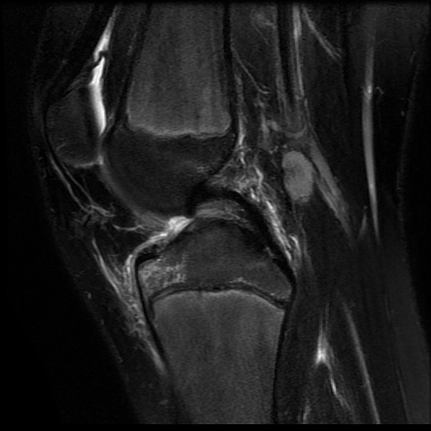
Bone bruises represent trabecular microfractures with haemorrhage and without a discrete fracture line or contour abnormality 4. They typically appear within 48 hours of injury and can persist for up to six months 3.
Aetiology
Most bone contusions are a result of a direct blow to the bone, traction from avulsion trauma, or load to a subchondral surface 1,2,5. Depending on where bone contusion is seen, the underlying trauma mechanism can be identified.
Complications
They can progress to osteochondritis dissecans 2.
Radiographic features
Plain radiograph
Plain radiography will not demonstrate cancellous features but can show regions of impaction, for example, a Hill-Sachs defect 5.
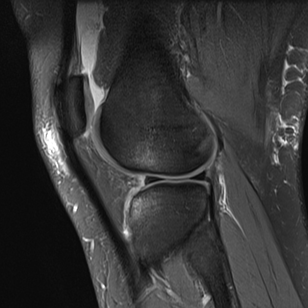
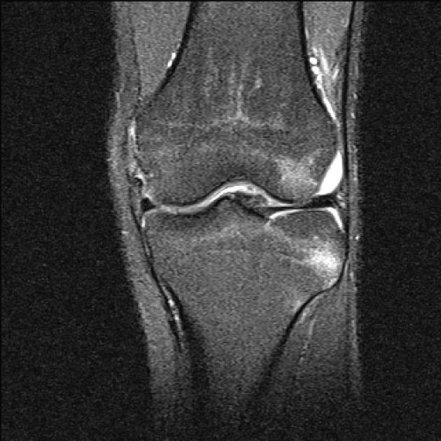
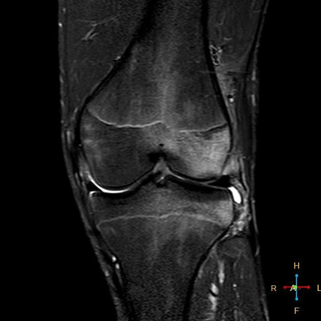
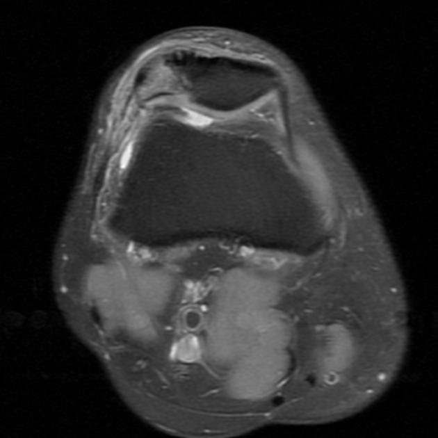
MRI
MRI is the modality of choice when investigating bone marrow. Bone (marrow) contusion is typically focal and ill-defined with the following signal characteristics 4:
- T1: focal hypointense area of bone marrow
- T2 fat-saturated: focal hyperintense area of bone marrow
ADVERTISEMENT: Supporters see fewer/no ads
See also
Quiz questions
References
- 1. Sanders T, Medynski M, Feller J, Lawhorn K. Bone Contusion Patterns of the Knee at MR Imaging: Footprint of the Mechanism of Injury. Radiographics. 2000;20 Spec No(suppl_1):S135-51. doi:10.1148/radiographics.20.suppl_1.g00oc19s135 - Pubmed
- 2. Helms CA. Fundamentals of Skeletal Radiology: Expert Consult: Online (Fundamentals of Radiology). Saunders. ISBN:B00F0ZS62Y. Read it at Google Books - Find it at Amazon
- 3. Harry Griffiths. Musculoskeletal Radiology. (2008) ISBN: 0849393906 - Google Books
- 4. Gorbachova T, Melenevsky Y, Cohen M, Cerniglia B. Osteochondral Lesions of the Knee: Differentiating the Most Common Entities at MRI. Radiographics. 2018;38(5):1478-95. doi:10.1148/rg.2018180044 - Pubmed
- 5. Palmer W, Bancroft L, Bonar F et al. Glossary of Terms for Musculoskeletal Radiology. Skeletal Radiol. 2020;49(Suppl 1):1-33. doi:10.1007/s00256-020-03465-1 - Pubmed
Incoming Links
- Patterns of bone bruise in knee injury
- Subchondral insufficiency fracture
- Posterior cruciate ligament tear
- Patellofemoral instability
- Trauma
- Anterior cruciate ligament tear
- Inferior shoulder dislocation
- Foot pain
- Rapidly destructive osteoarthritis of the hip
- Dorsal intercalated segment instability
- Focal periphyseal oedema zone
- Bone marrow oedema
- Acute vs chronic anterior cruciate ligament tears
- Contrecoup injury (knee)
- Patterns of bone bruise in knee injury
- Anterior cruciate ligament tear due to pivot shift
- Anterior cruciate ligament tear with bone bruises suggestive of pivot shift
- Knee hyperextension with typical bone bruises and posterior cruciate ligament tear
- Lateral patellar dislocation with characteristic bone bruises
- Focal periphyseal edema zones
- Lateral patellar dislocation with osteochondral injury
- Transient lateral patellar dislocation
- Scaphoid bone fracture
- Multiple carpal bone bruises
- Popliteus muscle injury and partial patellar tendon avulsion
- Anterior cruciate ligament tear - complete
- Isolated trapezoid fracture
- Avulsion fracture of the fibular head
- Nondisplaced incomplete intertrochanteric fracture
- Transient lateral patellar dislocation
- Annular pulley partial tear - thumb
- Lateral patellar dislocation injury with detached osteochondral fracture
- Transient patellar dislocation
- Intraarticular knee fractures
Related articles: Fractures
-
fracture
- terminology
- fracture location
- diaphyseal fracture
- metaphyseal fracture
- physeal fracture
- epiphyseal fracture
- fracture types
- avulsion fracture
- articular surface injuries
- complete fracture
- incomplete fracture
- infraction
- compound fracture
- pathological fracture
- stress fracture
- fracture displacement
- fracture location
- fracture healing
- skull fractures
-
facial fractures
- fractures involving a single facial buttress
- alveolar process fractures
- frontal sinus fracture
- isolated zygomatic arch fractures
- mandibular fracture
- nasal bone fracture
- orbital blow-out fracture
- paranasal sinus fractures
- complex fractures
- dental fractures
- fractures involving a single facial buttress
-
spinal fractures
- classification (AO Spine classification systems)
-
cervical spine fracture classification systems
- AO classification of upper cervical injuries
- AO classification of subaxial injuries
- Anderson and D'Alonzo classification (odontoid fracture)
- Roy-Camille classification (odontoid process fracture)
- Gehweiler classifcation (atlas fractures)
- Levine and Edwards classification (hangman fracture)
- Allen and Ferguson classification (subaxial spine injuries)
- subaxial cervical spine injury classification (SLIC)
- thoracolumbar spinal fracture classification systems
- three column concept of spinal fractures (Denis classification)
- classification of sacral fractures
-
cervical spine fracture classification systems
- spinal fractures by region
- spinal fracture types
- classification (AO Spine classification systems)
- rib fractures
- sternal fractures
-
upper limb fractures
- classification
- Rockwood classification (acromioclavicular joint injury)
- AO classification (clavicle fracture)
- Neer classification (clavicle fracture)
- Neer classification (proximal humeral fracture)
- AO classification (proximal humeral fracture)
- AO/OTA classification of distal humeral fractures
- Milch classification (lateral humeral condyle fracture)
- Weiss classification (lateral humeral condyle fracture)
- Bado classification of Monteggia fracture-dislocations (radius-ulna)
- Mason classification (radial head fracture)
- Frykman classification (distal radial fracture)
- Mayo classification (scaphoid fracture)
- Hintermann classification (gamekeeper's thumb)
- Eaton classification (volar plate avulsion injury)
- Keifhaber-Stern classification (volar plate avulsion injury)
- upper limb fractures by region
- shoulder
- clavicular fracture
-
scapular fracture
- acromion fracture
- coracoid process fracture
- glenoid fracture
- humeral head fracture
- proximal humeral fracture
- humeral neck fracture
- arm
- elbow
- forearm
- wrist
-
carpal bones
- scaphoid fracture
- lunate fracture
- capitate fracture
- triquetral fracture
- pisiform fracture
- hamate fracture
- trapezoid fracture
- trapezium fracture
- hand
- shoulder
- classification
- lower limb fractures
- classification by region
- pelvic fractures
- hip fractures
- Pipkin classification (femoral head fracture)
- Garden classification (hip fracture)
- American Academy of Orthopaedic Surgeons classification (periprosthetic hip fracture)
- Cooke and Newman classification (periprosthetic hip fracture)
- Johansson classification (periprosthetic hip fracture)
- Vancouver classification (periprosthetic hip fracture)
- femoral
- knee
- Schatzker classification (tibial plateau fracture)
- AO classification of distal femur fractures
- Meyers and McKeevers classification (anterior cruciate ligament avulsion fracture)
- tibia/fibula
- Watson-Jones classification (tibial tuberosity avulsion fracture)
- ankle
- foot
- Berndt and Harty classification (osteochondral lesions of the talus)
- Sanders CT classification (calcaneal fracture)
- Hawkins classification (talar neck fracture)
- Myerson classification (Lisfranc injury)
- Nunley-Vertullo classification (Lisfranc injury)
- pelvis and lower limb fractures by region
- pelvic fracture
- sacral fracture
- coccygeal fracture
-
hip
- acetabular fracture
- femoral head fracture
-
femoral neck fracture
- subcapital fracture
- transcervical fracture
- basicervical fracture
-
trochanteric fracture
- pertrochanteric fracture
- intertrochanteric fracture
- subtrochanteric fracture
- femur
- mid-shaft fracture
- bisphosphonate-related fracture
- distal femoral fracture
- knee
- avulsion fractures
- Segond fracture
- reverse Segond fracture
- anterior cruciate ligament avulsion fracture
- posterior cruciate ligament avulsion fracture
- arcuate complex avulsion fracture (arcuate sign)
- biceps femoris avulsion fracture
- iliotibial band avulsion fracture
- semimembranosus tendon avulsion fracture
- Stieda fracture (MCL avulsion fracture)
- patellar fracture
- tibial plateau fracture
- avulsion fractures
- leg
- tibial tuberosity avulsion fracture
- tibial shaft fracture
- fibular shaft fracture
- Maisonneuve fracture
- ankle
- foot
- tarsal bones
- metatarsal bones
- phalanges
- classification by region
- terminology


 Unable to process the form. Check for errors and try again.