Calcific tendinitis
Updates to Article Attributes
Calcific tendinitis (also known as calcific tendinopathy or tendonitis) is a self-limiting condition due to the deposition of calcium hydroxyapatite within tendons, usually of the rotator cuff. It is a common presentation of the hydroxyapatite crystal deposition disease (HADD).
Epidemiology
Typically this condition affects middle-aged patients between the ages of 30 and 60, with a slight predilection for women 2.
Clinical presentation
The condition passes through four stages 2:
- precalcific
- asymptomatic
- fibrocartilaginous metaplasia (see below)
- calcific or formative
- symptoms are variable from none to pain on movement
- resorptive
- most symptomatic
- pain due to extravasation of calcium hydroxyapatite into adjacent tissues, especially subacromial bursa, causing calcific bursitis
- pain typically lasts two weeks
- postcalcific
- variable symptomatology
- some restriction of movement common
- may last months
Pathology
Calcific tendinitis results from the deposition of calcium hydroxyapatite within the substance of a tendon and is thought to be due to decreased oxygen tension, leading to fibrocartilaginous metaplasia and secondary mineralisation 1.
Location
This condition most frequently affects the rotator cuff of the shoulder 1.
- supraspinatus: 80%
- infraspinatus: 15%
- subscapularis: 5%
- periarticular soft tissues in addition to tendons
- ligaments
- capsule
- bursae
However, the condition may occur anywhere in the body with the hip and knee joints being the 2nd and 3rd most common locations respectively 10,11.
Radiographic features
Plain radiograph
Calcific deposits are usually visualised as homogeneous hyperdensity with variable morphology, but typically globular/amorphous with smooth or ill-defined margins.
Ultrasound
Features of calcific tendinitis on ultrasound may include 7:
- a curvilinear/ovoid calcification with acoustic shadowing
- capsular soft tissue swelling
MRI
-
T1
- hypointense homogeneous signal
- adjacent tendon may be thickened
- some enhancement surrounding deposit may be seen
-
T2
- hypointense calcium deposits
- hyperintense signal may be present peripherally due to oedema
- hyperintense subacromial-subdeltoid bursal fluid
- T2*: calcifications may bloom
Treatment and prognosis
Controversial and difficult to measure due to the inherent variability of the symptoms and the self-limiting nature of the disease. Potential treatments include 2:
- oral analgesic/anti-inflammatory medication
- subacromial local anaesthetic/steroid injection
- aspiration of mineralised material
- ultrasound therapy
Differential diagnosis
In the shoulder consider:
- incidental calcification: seen in 2.5-20% of 'normal' healthy shoulders 1,2
- degenerative calcification
- seen in previously torn tendons
- generally smaller
- slightly older individuals
-
loose bodies
- associated chondral defect
- associated secondary osteoarthritis
-<p><strong>Calcific tendinitis</strong> (also known as <strong>calcific tendinopathy</strong> or <strong>tendonitis</strong>) is a <a href="/articles/self-limiting-2">self-limiting</a> condition due to the deposition of calcium hydroxyapatite within <a href="/articles/tendon">tendons</a>, usually of the <a href="/articles/rotator-cuff">rotator cuff</a>. It is a common presentation of the <a href="/articles/hydroxyapatite-deposition-disease">hydroxyapatite crystal deposition disease (HADD)</a>. </p><h4>Epidemiology</h4><p>Typically this condition affects middle-aged patients between the ages of 30 and 60, with a slight predilection for women <sup>2</sup>.</p><h4>Clinical presentation</h4><p>The condition passes through four stages <sup>2</sup>:</p><ol>-<li>precalcific<ul>-<li>asymptomatic</li>-<li>fibrocartilaginous metaplasia (see below)</li>-</ul>-</li>-<li>calcific or formative<ul><li>symptoms are variable from none to pain on movement</li></ul>-</li>-<li>resorptive<ul>-<li>most symptomatic</li>-<li>pain due to extravasation of calcium hydroxyapatite into adjacent tissues, especially <a href="/articles/subacromial-subdeltoid-bursa">subacromial bursa</a>, causing <a href="/articles/calcific-bursitis">calcific bursitis</a>-</li>-<li>pain typically lasts two weeks</li>-</ul>-</li>-<li>postcalcific<ul>-<li>variable symptomatology</li>-<li>some restriction of movement common</li>-<li>may last months</li>-</ul>-</li>-</ol><h4>Pathology</h4><p>Calcific tendinitis results from the deposition of calcium hydroxyapatite within the substance of a tendon and is thought to be due to decreased oxygen tension, leading to fibrocartilaginous metaplasia and secondary mineralisation <sup>1</sup>.</p><h5>Location</h5><p>This condition most frequently affects the <a href="/articles/rotator-cuff">rotator cuff</a> of the shoulder <sup>1</sup>.</p><ul>-<li>-<a href="/articles/supraspinatus-muscle-1">supraspinatus</a>: 80%</li>-<li>-<a href="/articles/infraspinatus-muscle-1">infraspinatus</a>: 15%</li>-<li>-<a href="/articles/subscapularis-muscle-2">subscapularis</a>: 5%</li>-<li>periarticular soft tissues in addition to tendons</li>-<li>ligaments</li>-<li>capsule</li>-<li>bursae</li>-</ul><p>However, the condition may occur anywhere in the body with the <a href="/articles/hip-joint-1">hip</a> and <a href="/articles/knee-joint-1">knee</a> joints being the 2<sup>nd</sup> and 3<sup>rd</sup> most common locations respectively <sup>10,11</sup>.</p><h4>Radiographic features</h4><h5>Plain radiograph</h5><p>Calcific deposits are usually visualised as homogeneous hyperdensity with variable morphology, but typically globular/amorphous with smooth or ill-defined margins.</p><h5>Ultrasound</h5><p>Features of calcific tendinitis on ultrasound may include <sup>7</sup>:</p><ul>-<li>a curvilinear/ovoid calcification with acoustic shadowing</li>-<li>capsular soft tissue swelling</li>-</ul><h5>MRI</h5><ul>-<li>-<strong>T1</strong><ul>-<li>hypointense homogeneous signal</li>-<li>adjacent tendon may be thickened</li>-<li>some enhancement surrounding deposit may be seen</li>-</ul>-</li>-<li>-<strong>T2</strong><ul>-<li>hypointense calcium deposits</li>-<li>hyperintense signal may be present peripherally due to oedema</li>-<li>hyperintense subacromial-subdeltoid bursal fluid</li>-</ul>-</li>-<li>-<strong>T2</strong>*: calcifications may bloom</li>-</ul><h4>Treatment and prognosis</h4><p>Controversial and difficult to measure due to the inherent variability of the symptoms and the self-limiting nature of the disease. Potential treatments include <sup>2</sup>:</p><ul>-<li>oral analgesic/anti-inflammatory medication</li>-<li>subacromial local anaesthetic/steroid injection</li>-<li>aspiration of mineralised material</li>-<li>ultrasound therapy</li>-</ul><h4>Differential diagnosis</h4><p>In the shoulder consider:</p><ul>-<li>incidental calcification: seen in 2.5-20% of 'normal' healthy shoulders <sup>1,2</sup>-</li>-<li>degenerative calcification<ul>-<li>seen in previously torn tendons</li>-<li>generally smaller</li>-<li>slightly older individuals</li>-</ul>-</li>-<li>-<a href="/articles/intra-articular-loose-bodies-2">loose bodies</a><ul>-<li>associated chondral defect</li>-<li>associated secondary osteoarthritis</li>-</ul>-</li>- +<p><strong>Calcific tendinitis</strong> (also known as <strong>calcific tendinopathy</strong> or <strong>tendonitis</strong>) is a <a href="/articles/self-limiting-2">self-limiting</a> condition due to the deposition of calcium hydroxyapatite within <a href="/articles/tendon">tendons</a>, usually of the <a href="/articles/rotator-cuff">rotator cuff</a>. It is a common presentation of the <a href="/articles/hydroxyapatite-deposition-disease">hydroxyapatite crystal deposition disease (HADD)</a>. </p><h4>Epidemiology</h4><p>Typically this condition affects middle-aged patients between the ages of 30 and 60, with a slight predilection for women <sup>2</sup>.</p><h4>Clinical presentation</h4><p>The condition passes through four stages <sup>2</sup>:</p><ol>
- +<li>precalcific<ul>
- +<li>asymptomatic</li>
- +<li>fibrocartilaginous metaplasia (see below)</li>
- +</ul>
- +</li>
- +<li>calcific or formative<ul><li>symptoms are variable from none to pain on movement</li></ul>
- +</li>
- +<li>resorptive<ul>
- +<li>most symptomatic</li>
- +<li>pain due to extravasation of calcium hydroxyapatite into adjacent tissues, especially <a href="/articles/subacromial-subdeltoid-bursa">subacromial bursa</a>, causing <a href="/articles/calcific-bursitis">calcific bursitis</a>
- +</li>
- +<li>pain typically lasts two weeks</li>
- +</ul>
- +</li>
- +<li>postcalcific<ul>
- +<li>variable symptomatology</li>
- +<li>some restriction of movement common</li>
- +<li>may last months</li>
- +</ul>
- +</li>
- +</ol><h4>Pathology</h4><p>Calcific tendinitis results from the deposition of calcium hydroxyapatite within the substance of a tendon and is thought to be due to decreased oxygen tension, leading to fibrocartilaginous metaplasia and secondary mineralisation <sup>1</sup>.</p><h5>Location</h5><p>This condition most frequently affects the <a href="/articles/rotator-cuff">rotator cuff</a> of the shoulder <sup>1</sup>.</p><ul>
- +<li>
- +<a href="/articles/supraspinatus-muscle-1">supraspinatus</a>: 80%</li>
- +<li>
- +<a href="/articles/infraspinatus-muscle-1">infraspinatus</a>: 15%</li>
- +<li>
- +<a href="/articles/subscapularis-muscle-2">subscapularis</a>: 5%</li>
- +<li>periarticular soft tissues in addition to tendons</li>
- +<li>ligaments</li>
- +<li>capsule</li>
- +<li>bursae</li>
- +</ul><p>However, the condition may occur anywhere in the body with the <a href="/articles/hip-joint-1">hip</a> and <a href="/articles/knee-joint-1">knee</a> joints being the 2<sup>nd</sup> and 3<sup>rd</sup> most common locations respectively <sup>10,11</sup>.</p><h4>Radiographic features</h4><h5>Plain radiograph</h5><p>Calcific deposits are usually visualised as homogeneous hyperdensity with variable morphology, but typically globular/amorphous with smooth or ill-defined margins.</p><h5>Ultrasound</h5><p>Features of calcific tendinitis on ultrasound may include <sup>7</sup>:</p><ul>
- +<li>a curvilinear/ovoid calcification with acoustic shadowing</li>
- +<li>capsular soft tissue swelling</li>
- +</ul><h5>MRI</h5><ul>
- +<li>
- +<strong>T1</strong><ul>
- +<li>hypointense homogeneous signal</li>
- +<li>adjacent tendon may be thickened</li>
- +<li>some enhancement surrounding deposit may be seen</li>
- +</ul>
- +</li>
- +<li>
- +<strong>T2</strong><ul>
- +<li>hypointense calcium deposits</li>
- +<li>hyperintense signal may be present peripherally due to oedema</li>
- +<li>hyperintense subacromial-subdeltoid bursal fluid</li>
- +</ul>
- +</li>
- +<li>
- +<strong>T2</strong>*: calcifications may bloom</li>
- +</ul><h4>Treatment and prognosis</h4><p>Controversial and difficult to measure due to the inherent variability of the symptoms and the self-limiting nature of the disease. Potential treatments include <sup>2</sup>:</p><ul>
- +<li>oral analgesic/anti-inflammatory medication</li>
- +<li>subacromial local anaesthetic/steroid injection</li>
- +<li>aspiration of mineralised material</li>
- +<li>ultrasound therapy</li>
- +</ul><h4>Differential diagnosis</h4><p>In the shoulder consider:</p><ul>
- +<li>incidental calcification: seen in 2.5-20% of 'normal' healthy shoulders <sup>1,2</sup>
- +</li>
- +<li>degenerative calcification<ul>
- +<li>seen in previously torn tendons</li>
- +<li>generally smaller</li>
- +<li>slightly older individuals</li>
- +</ul>
- +</li>
- +<li>
- +<a href="/articles/intra-articular-loose-bodies-2">loose bodies</a><ul>
- +<li>associated chondral defect</li>
- +<li>associated secondary osteoarthritis</li>
- +</ul>
- +</li>
Image 33 CT (bone window) ( create )
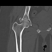
Image 34 CT (bone window) ( create )
