
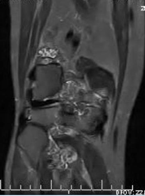
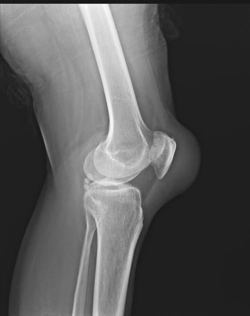
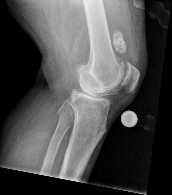
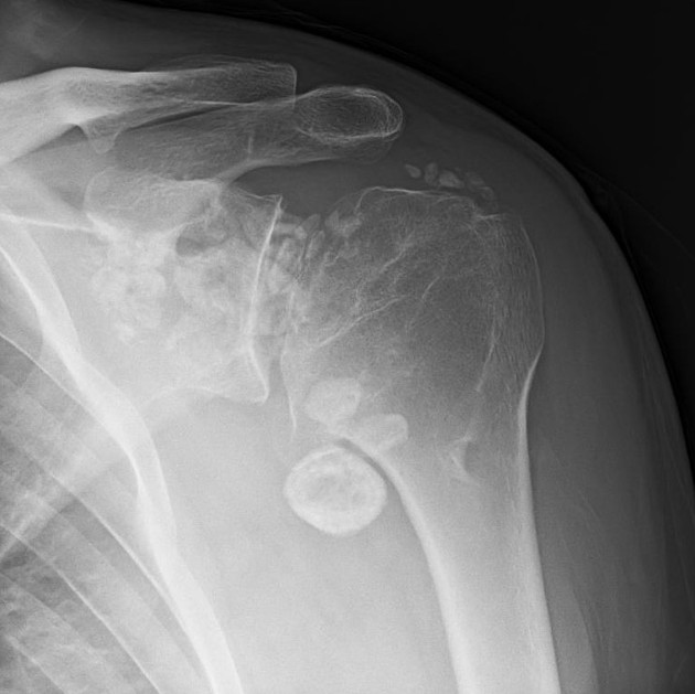
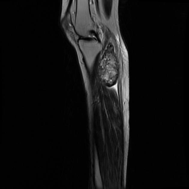
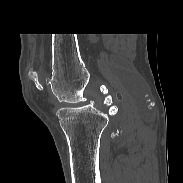
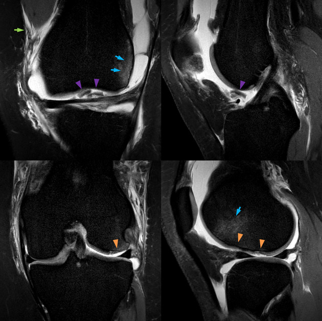
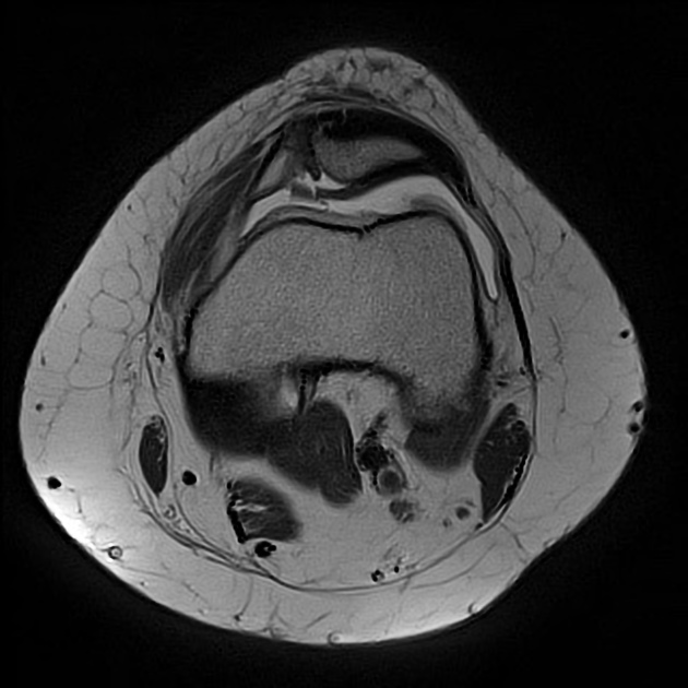
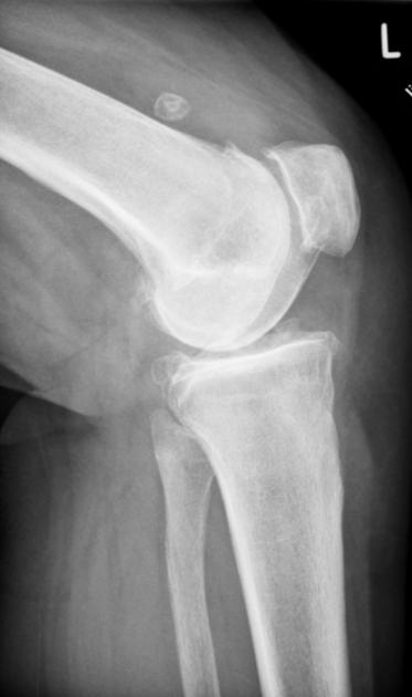
Intra-articular loose bodies can result from a variety of pathological processes.
On this page:
Terminology
The use of the term "loose" is frowned upon by some authors because the fragments do not necessarily move around in the joint ref - the term intra-articular body or fragment is a safer alternative. Historically they were often called joint mice.
Clinical presentation
Patients may be entirely asymptomatic or complain of pain, clicking and locking, depending on the location and mobility of the fragment as well as any associated secondary degenerative disease, and symptoms from the underlying cause.
Pathology
Intra-articular bodies are composed of cartilage or cartilage and bone and result from any process that leads to disruption of the articular surface. They derive nutrition from synovial fluid and contain any of the cells of bone or cartilage. The surface cells form more cartilaginous layers, so enlarging the body over time. Deeper cells receive less nutrition resulting in cell death and calcification 2.
Etiology
A wide range of conditions can lead to the development of intra-articular loose bodies:
osteoarthritis or severe degenerative disease
meniscal fragmentation and its calcification
fragmented/fractured osteophytes 6


 Unable to process the form. Check for errors and try again.