
Cardiac rhabdomyomas are a type of benign myocardial tumour and are considered the most common fetal cardiac tumour. They have a strong association with tuberous sclerosis.
On this page:
Epidemiology
Cardiac rhabdomyomas are often multiple and can represent up to 90% of cardiac tumours in the paediatric population 1. The majority are diagnosed before the age of 1 year. The estimated incidence is at ~1 in 20,000 births 8.
Associations
there is a well-known association with tuberous sclerosis, with >50% of all cardiac rhabdomyomas found in patients with later confirmed tuberous sclerosis 1,2
Clinical presentation
The majority of cardiac rhabdomyomas are asymptomatic although there can be a broad clinical spectrum. Occasionally, they may present with left ventricular outflow tract obstruction or refractory arrhythmias.
Pathology
It is a hamartomatous lesion consisting of cardiac muscle tissue (derived from embryonal myoblasts).
Location
They may arise anywhere in the myocardium but are more common in the ventricles (may involve the left ventricle the most) 6.
Macroscopic appearance
Macroscopically, they appear as yellow-tan solid, circumscribed, unencapsulated lesions.
Microscopic appearance
Microscopically, a characteristic spider cell is seen which is a large clear cell with cytoplasmic strands composed of glycogen extending to the plasma membrane.
Radiographic features
Ultrasound/echocardiography
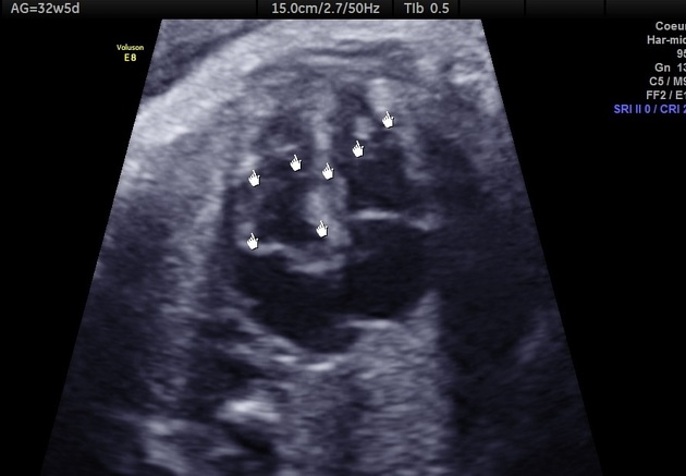
May be seen as one or more solid hyperechoic masses located in relation to the myocardium. Small lesions can mimic diffuse myocardial thickening. They frequently occur in relation to the ventricles. The size of a lesion detected in utero may range from 10-50 mm 6.
MRI
T1: isointense to adjacent myocardium
T2: hyperintense to adjacent myocardium
Treatment and prognosis
In most cases, no treatment is required, and these lesions regress spontaneously. Patients with left ventricular outflow tract obstruction or refractory arrhythmias respond well to surgical excision. The overall prognosis is dependent on the number, size and location of the lesions as well as the presence or absence of associated anomalies.
Complications
development of cardiac arrhythmias
-
intracavitary growth may cause
ventricular outflow tract obstruction
valvular compromise
disruption of intracardiac blood flows leading to congestive heart failure and hydrops
Differential diagnosis
For the in-utero sonographic appearance of diffuse myocardial thickening, consider:


 Unable to process the form. Check for errors and try again.