Disclosures
- updated 17 Aug 2022:
Nothing to disclose
Updates to Article Attributes
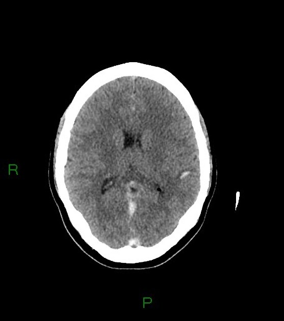
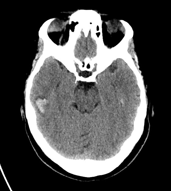
The cashew nut sign is a radiological sign described in juxtacortical intracerebral haemorrhages due to cerebral venous thrombosis, typically on CT.
The sign describes a small (<20 mm), concave-shaped intracerebral haemorrhage in the juxtacortical white matter, often near the bottom of a cortical sulcus, which resembles a cashew nut 1-3. Presence of juxtacortical intracerebral haemorrhages has been found to be very specific (98%) for intracerebral haemorrhage due to cerebral venous thrombosis, typically of the of the superior sagittal sinus, but not very sensitive (26%) 1,2.
It is thought that this unique cashew nut shape and juxtacortical location of haemorrhage is due to involvement of arcuate segments of the subcortical veins, which run parallel to subcortical U-fibres 2,3. These subcortical veins drain into cortical veins and then venous sinuses and thus are affected by cerebral venous thrombosis 2,3.
-<p>The <strong>cashew nut sign</strong> is a radiological sign described in juxtacortical <a href="/articles/intracerebral-haemorrhage">intracerebral haemorrhages</a> due to <a href="/articles/cerebral-venous-thrombosis">cerebral venous thrombosis</a>, typically on CT.</p><p>The sign describes a small (<20 mm), concave-shaped <a href="/articles/intracerebral-haemorrhage">intracerebral haemorrhage</a> in the juxtacortical white matter, often near the bottom of a cortical sulcus, which resembles a cashew nut <sup>1-3</sup>. Presence of juxtacortical <a href="/articles/intracerebral-haemorrhage">intracerebral haemorrhages</a> has been found to be very specific (98%) for <a href="/articles/intracerebral-haemorrhage">intracerebral haemorrhage</a> due to <a href="/articles/cerebral-venous-thrombosis">cerebral venous thrombosis</a>, typically of the of the <a href="/articles/superior-sagittal-sinus">superior sagittal sinus</a>, but not very sensitive (26%) <sup>1,2</sup>.</p><p>It is thought that this unique cashew nut shape and juxtacortical location of haemorrhage is due to involvement of arcuate segments of the subcortical veins which run parallel to <a href="/articles/subcortical-u-fibres-3">subcortical U-fibres</a> <sup>2,3</sup>. These subcortical veins drain into cortical veins and then venous sinuses and thus are affected by <a href="/articles/cerebral-venous-thrombosis">cerebral venous thrombosis</a> <sup>2,3</sup>. </p>- +<p>The <strong>cashew nut sign</strong> is a radiological sign described in juxtacortical <a href="/articles/intracerebral-haemorrhage">intracerebral haemorrhages</a> due to <a href="/articles/cerebral-venous-thrombosis">cerebral venous thrombosis</a>, typically on CT.</p><p>The sign describes a small (<20 mm), concave-shaped <a href="/articles/intracerebral-haemorrhage">intracerebral haemorrhage</a> in the juxtacortical white matter, often near the bottom of a cortical sulcus, which resembles a cashew nut <sup>1-3</sup>. Presence of juxtacortical <a href="/articles/intracerebral-haemorrhage">intracerebral haemorrhages</a> has been found to be very specific (98%) for <a href="/articles/intracerebral-haemorrhage">intracerebral haemorrhage</a> due to <a href="/articles/cerebral-venous-thrombosis">cerebral venous thrombosis</a>, typically of the <a href="/articles/superior-sagittal-sinus">superior sagittal sinus</a>, but not very sensitive (26%) <sup>1,2</sup>.</p><p>It is thought that this unique cashew nut shape and juxtacortical location of haemorrhage is due to involvement of arcuate segments of the subcortical veins, which run parallel to <a href="/articles/subcortical-u-fibres-3">subcortical U-fibres</a> <sup>2,3</sup>. These subcortical veins drain into cortical veins and then venous sinuses and thus are affected by <a href="/articles/cerebral-venous-thrombosis">cerebral venous thrombosis</a> <sup>2,3</sup>. </p>


 Unable to process the form. Check for errors and try again.