
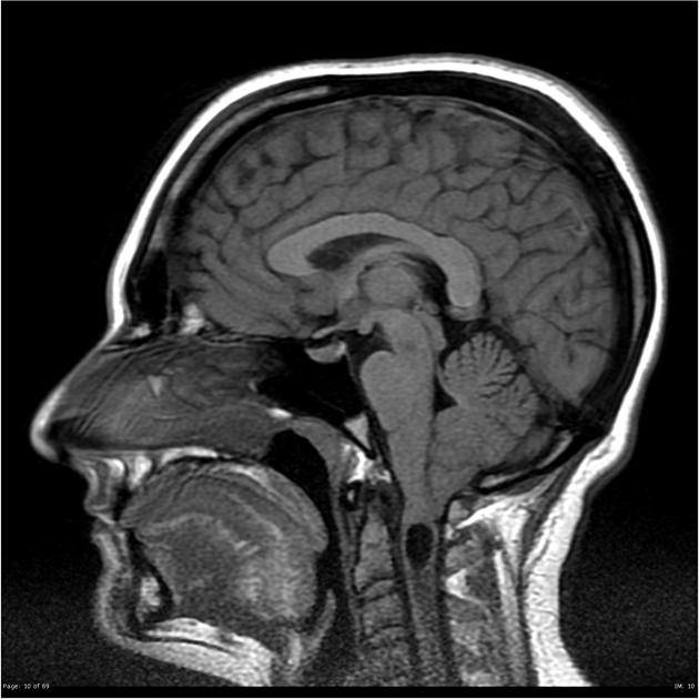
Chiari malformations are a group of structural conditions characterised by congenital caudal 'displacement' of the cerebellar tonsils below the foramen magnum, often with associated caudal displacement of brainstem.
On this page:
Terminology
The description of 'malformation' may not be accurate for Chiari 0, 0.5, 1 and 1.5 because there is evidence that these are defects acquired after birth 8. Thus, the term 'developmental alteration' or 'deformation' has been suggested for these entities instead 8,9. Chiari 2 to 5, on the other hand, are all considered congenital malformations 8.
Classification
The initial description by Hans Chiari in 1891, based on autopsy observations of children, included three types (Chiari 1, 2 and 3). A few years later (see below) he described a fourth type (Chiari 4). Additional types have subsequently been described, raising the total number to 9, although how these conditions are related to each other remains a topic of debate 1,9.
Some have advocated that these nine types can be divided into two broad groups: primary Chiari malformation (Chiari 0 to 2) and complex Chiari malformations (Chiari 3 to 5) although no clear consensus exists 9.
Additionally, there is increasing support for the view that Chiari 0, 0.5 and 1 represent a continuum and that relying on cerebellar tonsilar position is creating confusion 9.
Be that as it may, the 9 Chiari types are as follows 1,8,9:
-
Chiari 0
obliteration of the cisterna magna and/or small posterior fossa
no cerebellar tonsillar descent (<5 mm caudal descent in relation to the foramen magnum); but
presence of a syrinx and presence of typical Chiari symptoms
infrequently used and confusing term to be avoided
based on reports of syrinx improvement after posterior fossa decompression
-
Chiari 0.5
no cerebellar tonsillar descent (<5 mm caudal descent in relation to the foramen magnum); but
tonsillar position is ventral to a line bisecting the medulla
-
most common
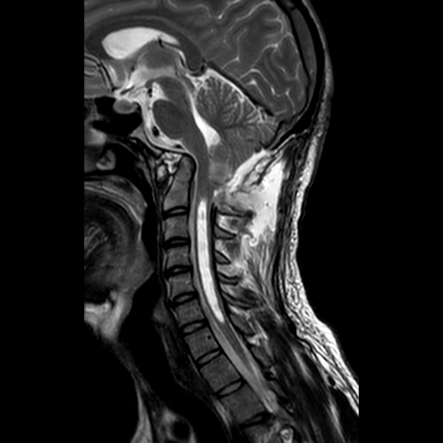
cerebellar tonsillar herniation (>5 mm caudal herniation in relation to the foramen magnum) alone
there may or may not be an associated syrinx
not associated with nueral tube defects
-
cerebellar tonsillar herniation (>5 mm caudal herniation in relation to the foramen magnum); with
herniation of the obex inferior to the foramen magnum
not associated with nueral tube defects
-
cerebellar tonsillar (>5 mm caudal herniation), brainstem, and fourth ventricle herniation in relation to the foramen magnum; with
associated lumbosacral myelomeningocele
-
similar features to Chiari 2; but with
an occipital and/or high cervical encephalocele instead of a lumbosacral myelomeningocele
-
Chiari 3.5
similar features to Chiari 3; but with
an occipitocervical myeloencephalocele and foregut communication
-
Chiari 4
severe cerebellar hypoplasia without displacement of the cerebellum through the foramen magnum
associated with an occipital encephalocele
probably a variation of cerebellar hypoplasia
-
Chiari 5
absent cerebellum with myelomeningocele
herniation of the occipital lobe through the foramen magnum
Treatment and prognosis
The main objective of surgery in Chiari malformation is to improve CSF flow across the foramen magnum and surrounding the brainstem, reduce the extent of the syrinx, and reduce pressure on the brainstem 7.
History and etymology
A number of individual descriptions that in retrospect represent Chairi malformations date back to 1829 9.
Chiari malformations type 1 to 3 were first systematically described by Hans Chiari (1851-1914), an Austrian pathologist, in 1891 3. Four years later in 1895 he described a fourth type (Chiari 4) 10. In these papers, Chiari also credited Julius Arnold (1835-1915), Professor of Anatomy at Heidelberg, on the grounds of a previous publication of a case believed by Arnold to be of a Chiari 2 malformation. It appears that this is not actually the case, and as such the term Arnold-Chiari to denote Chiari 2 malformations is no longer advocated 4.
See also
incidental tonsillar ectopia


 Unable to process the form. Check for errors and try again.