
Cholecystocolonic fistulas are most commonly a rare late complication of gallstone disease, resulting from an abnormal communication between the gallbladder and the colon. It is the second most common cholecystoenteric fistula after cholecystoduodenal fistulas 1.
On this page:
Clinical presentation
These may be completely asymptomatic 2. Patient presentation is variable and may include abdominal pain, chronic diarrhoea, fever, nausea and vomiting, steatorrhoea, jaundice, and/or weight loss 1,3.
Complications
These fistulas can lead to pneumobilia and liver abscesses. In the case of stone impaction in the colon, it may cause a large bowel obstruction.
Pathology
Aetiology
Causes of cholecystocolonic fistulas include 2,3:
gallstones / cholecystitis (most common)
malignancy
trauma
Radiographic features
Cholecystocolonic fistulas are rarely diagnosed before surgery 1,3. ERCP is considered the most accurate modality 2,3.
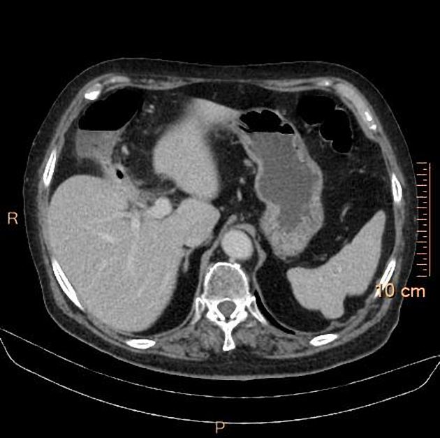
CT/MRI
There may be gallbladder wall thickening +/- intraluminal air. Pneumobilia may be present. The gallbladder may be adherent to the right side of the colon with a fistula sometimes visualised. Gallstone(s) may be visible within the fistula or within the colonic lumen.
Treatment and prognosis
No consensus exists with regards to optimal treatment. Surgical treatment can vary from minimally invasive procedures to extensive resection.


 Unable to process the form. Check for errors and try again.