
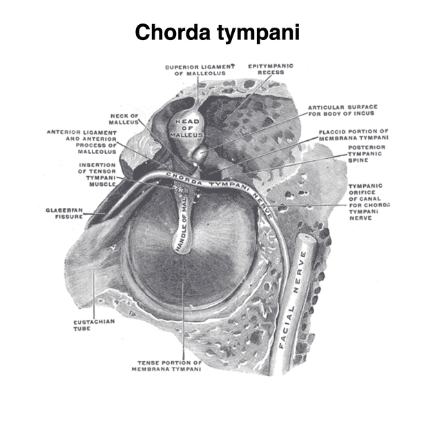
Chorda tympani
Citation, DOI, disclosures and article data
At the time the article was created Charlie Chia-Tsong Hsu had no recorded disclosures.
View Charlie Chia-Tsong Hsu's current disclosuresAt the time the article was last revised Craig Hacking had the following disclosures:
- Philips Australia, Paid speaker at Philips Spectral CT events (ongoing)
These were assessed during peer review and were determined to not be relevant to the changes that were made.
View Craig Hacking's current disclosures- Chorda tympani nerve
- Chordae tympani
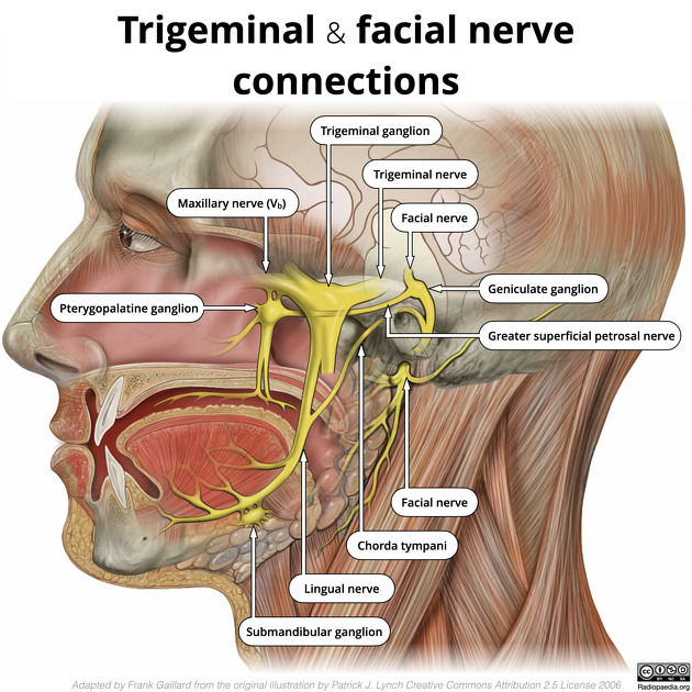
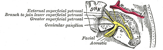
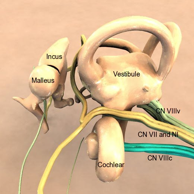
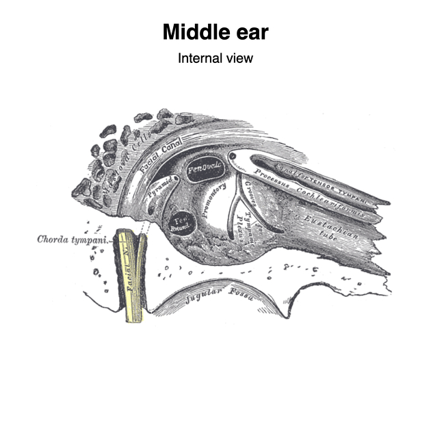
The chorda tympani is a nerve that arises from the mastoid segment of the facial nerve, carrying afferent special sensation from the anterior two-thirds of the tongue via the lingual nerve, as well as efferent parasympathetic secretomotor innervation to the submandibular and sublingual glands.
Gross anatomy
After branching off from the facial nerve, the chorda tympani courses through the temporal bone before joining the lingual nerve 2:
branch from mastoid segment of facial nerve: the branching site can be variable from either the proximal, mid- or distal mastoid segment of facial nerve; occasionally the chorda tympani can even branch off from the facial nerve after exiting the stylomastoid foramen
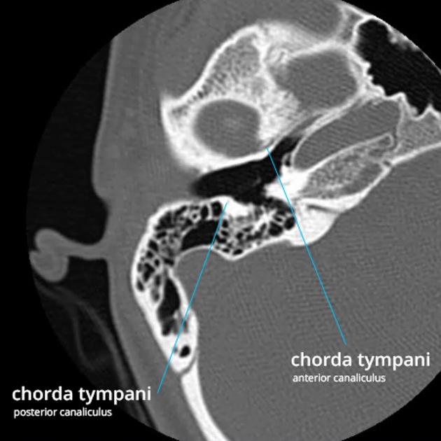
posterior canaliculus: after originating, the chorda tympani courses superiorly back into the tympanic cavity to the level of the manubrium/neck of malleus. The distance of ascent is variable, depending on the initial branching pattern from the mastoid segment of facial nerve
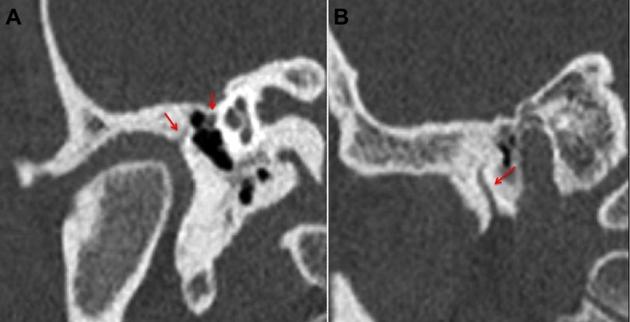
tympanic segment is the segment of chorda tympani as it traverses the middle ear cavity between the malleus and incus in a posteroanterior direction; not visible with current imaging techniques
anterior canaliculus: at the anterior wall of the tympanic cavity, it enters petrotympanic fissure before finally exiting the temporal bone
the chorda tympani exits the petrotympanic fissure medial to to the temporomandibular joint, within the infratemporal fossa. It then travels inferiorly to join the lingual nerve approximately 2 cm below the skull base
References
- 1. Susan Standring. Gray's Anatomy. (2008) ISBN: 9780443066849 - Google Books
- 2. Singh D, Hsu C, Kwan G, Bhuta S, Skalski M, Jones R. High Resolution CT Study of the Chorda Tympani Nerve and Normal Anatomical Variation. Jpn J Radiol. 2015;33(5):279-86. doi:10.1007/s11604-015-0417-2 - Pubmed
Incoming Links
- Middle ear tumours
- Lingual nerve
- Anterior tympanic artery
- Retrotympanum
- Tongue
- Parasympathetic nervous system
- Nervus intermedius
- Middle ear
- Mesotympanum
- Petrotympanic fissure
- Infratemporal fossa
- Greater wing of sphenoid
- Sublingual gland
- Tympanic membrane
- Facial nerve
- Submandibular ganglion
- Submandibular gland
- Nervus intermedius schwannoma
Related articles: Anatomy: Head and neck
- skeleton of the head and neck
-
cranial vault
- scalp (mnemonic)
- fontanelle
-
sutures
- calvarial
- facial
- frontozygomatic suture
- frontomaxillary suture
- frontolacrimal suture
- frontonasal suture
- temporozygomatic suture
- zygomaticomaxillary suture
- parietotemporal suture (parietomastoid suture)
- occipitotemporal suture (occipitomastoid suture)
- sphenofrontal suture
- sphenozygomatic suture
- spheno-occipital suture (not a true suture)
- lacrimomaxillary suture
- nasomaxillary suture
- internasal suture
- basal/internal
- skull landmarks
- frontal bone
- temporal bone
- parietal bone
- occipital bone
- skull base (foramina)
-
facial bones
- midline single bones
- paired bilateral bones
- cervical spine
- hyoid bone
- laryngeal cartilages
-
cranial vault
- muscles of the head and neck
- muscles of the tongue (mnemonic)
- muscles of mastication
-
facial muscles
- epicranius muscle
- circumorbital and palpebral muscles
- nasal muscles
-
buccolabial muscles
- elevators, retractors and evertors of the upper lip
- levator labii superioris alaeque nasalis muscle
- levator labii superioris muscle
- zygomaticus major muscle
- zygomaticus minor muscle
- levator anguli oris muscle
- malaris muscle
- risorius muscle
- depressors, retractors and evertors of the lower lip
- depressor labii inferioris muscle
- depressor anguli oris muscle
- mentalis muscle
- compound sphincter
-
orbicularis oris muscle
- incisivus labii superioris muscle
- incisivus labii inferioris muscle
-
orbicularis oris muscle
- muscle of mastication
- modiolus
- elevators, retractors and evertors of the upper lip
- muscles of the middle ear
- orbital muscles
- muscles of the soft palate
- pharyngeal muscles
- suprahyoid muscles
- infrahyoid muscles
- intrinsic muscles of the larynx
- muscles of the neck
- platysma muscle
- longus colli muscle
- longus capitis muscle
- scalenus anterior muscle
- scalenus medius muscle
- scalenus posterior muscle
- scalenus pleuralis muscle
- sternocleidomastoid muscle
-
suboccipital muscles
- rectus capitis posterior major muscle
- rectus capitis posterior minor muscle
- obliquus capitis superior muscle
- obliquus capitis inferior muscle
- accessory muscles of the neck
- deep cervical fascia
-
deep spaces of the neck
- anterior cervical space
- buccal space
- carotid space
- danger space
- deep cervical fascia
- infratemporal fossa
- masticator space
- parapharyngeal space
- stylomandibular tunnel
- parotid space
- pharyngeal (superficial) mucosal space
- perivertebral space
- posterior cervical space
- pterygopalatine fossa
- retropharyngeal space
- suprasternal space (of Burns)
- visceral space
- surgical triangles of the neck
- orbit
- ear
- paranasal sinuses
- upper respiratory tract
- viscera of the neck
- blood supply of the head and neck
-
arterial supply
-
common carotid artery
- carotid body
- carotid bifurcation
- subclavian artery
- variants
-
common carotid artery
- venous drainage
-
arterial supply
- innervation of the head and neck
-
cranial nerves
- olfactory nerve (CN I)
- optic nerve (CN II)
- oculomotor nerve (CN III)
- trochlear nerve (CN IV)
-
trigeminal nerve (CN V) (mnemonic)
- trigeminal ganglion
- ophthalmic division
- maxillary division
- mandibular division
- abducens nerve (CN VI)
- facial nerve (CN VII)
-
vestibulocochlear nerve (CN VIII)
- vestibular ganglion (Scarpa's ganglion)
- glossopharyngeal nerve (CN IX)
- vagus nerve (CN X)
- (spinal) accessory nerve (CN XI)
- hypoglossal nerve (CN XII)
- parasympathetic ganglia of the head and neck
- cervical sympathetic ganglia
- greater occipital nerve
- third occipital nerve
-
cervical plexus
- muscular branches
- longus capitis
- longus colli
- scalenes
- geniohyoid
- thyrohyoid
-
ansa cervicalis
- omohyoid (superior and inferior bellies separately)
- sternothyroid
- sternohyoid
- phrenic nerve
- contribution to the accessory nerve (CN XI)
- cutaneous branches
- muscular branches
- brachial plexus
- pharyngeal plexus
-
cranial nerves
- lymphatic drainage of the head and neck
- embryological development of the head and neck


 Unable to process the form. Check for errors and try again.