Chronic myocarditis is a prolonged or ongoing myocardial inflammation in the setting of non-dilated or mildly dilated cardiomyopathy 1-5. There have been significant differences concerning the exact definition of the concept as well as the time interval after the onset of symptoms, with the latter ranging from 14 days to 3 months after onset in previous definitions 1-4, and with subsequent definitions suggesting >30 days after onset 1,2.
In addition to the temporal definition, there is a pathological one, which characterizes the condition histologically as an inflammatory infiltrate of the myocardium with myocardial fibrosis and distinguishes the term between the usual chronic or chronic persistent form and chronic active myocarditis by the presence of cardiomyocyte injury or necrosis in one part of the world 1,2. The first form, chronic myocarditis, is also seen as a transitional stage between acute myocarditis and chronic inflammatory cardiomyopathy 1,2.
On this page:
Terminology
The term chronic myocarditis features some overlap with chronic inflammatory cardiomyopathy as both terms use a period of >30 days for their definition. However, the latter is also characterized by systolic dysfunction (see respective article). ‘Chronic active myocarditis’ and ‘chronic persistent myocarditis’ are historically based terms 6 and only the first is precisely defined within the Japanese guidelines 2.
In addition, the chronic myocarditis features some overlap with the terms ‘subacute myocarditis’ and chronic inflammatory cardiomyopathy. The term ‘subacute myocarditis’ is no longer encouraged 1.
Epidemiology
Associations
Chronic myocarditis has been associated with the following 5-8:
dilated cardiomyopathy (variable prevalence up to 50% or more 5)
Diagnosis
Diagnosis of myocarditis is made by a combination of clinical and imaging findings as well as pathological evidence of an ongoing inflammatory infiltrate within the myocardium. As invasive biopsy rates steadily decline, establishing the diagnosis has shifted to a combination of typical clinical findings and characteristic criteria on cardiac MRI 9. For the diagnosis of chronic myocarditis, the temporal definition >30 days after onset is of importance 1,2.
Clinical presentation
Typically there is a history of acute myocarditis or symptom onset >30 days prior. The initial complaints and the subsequent clinical course can be quite variable ranging from subclinical asymptomatic course to recurrent or persistent symptoms of heart failure and/or arrhythmia 2. Signs and symptoms include dyspnea, fatigue, peripheral edema, palpitations, syncope etc. 2.
Persistent increase in the blood high-sensitivity cardiac troponin levels can indicate chronic active myocarditis 2.
Pathology
Chronic myocarditis is characterized by the following 1-3:
ongoing inflammatory cell infiltration of the myocardium 6,7
histological evidence of fibrosis and/or cardiomyopathy-like changes within the cardiomyocytes 2,7
A setting with evidence of cardiomyocyte injury, necrosis or cell degeneration associated with an infiltration of inflammatory cells at the cardiomyocyte circumference in the myocardium indicates chronic active myocarditis as per the Japanese 2023 JCS guidelines 2.
Immunophenotype
An immunohistochemistry stain positive for tenascin C (4C8) 2,7, or the presence of ≥24 CD3+ T cells/mm2 in the myocardial tissue can indicate chronic active myocarditis 1.
Radiographic features
MRI
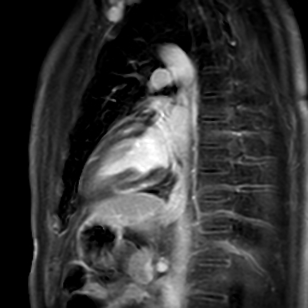
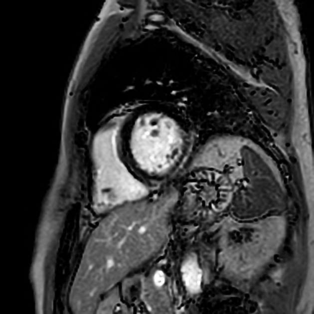
The sensitivity of cardiac MRI in the diagnosis of chronic myocarditis is significantly lower than in acute myocarditis. In particular, myocardial edema is less common and has been observed in only 25-30% of cases compared with >80% in the acute setting 11.
The presence of late gadolinium enhancement (LGE) has been well documented in chronic myocarditis 12-16 and is also of significant prognostic value especially if not accompanied by myocardial edema 15,16. However, late gadolinium enhancement is also non-specific and may be seen in other non-inflammatory cardiomyopathies 4. Patterns of late gadolinium enhancement are similar to those seen in acute myocarditis and include subepicardial and midwall patterns, the latter of which appears to be slightly more common in the chronic form 4. An increase in the late gadolinium enhancement extent and septal enhancement features a worse prognosis 15-17.
Native T1 and ECV may indicate myocardial edema, myocardial injury and myocardial fibrosis and may be elevated in the setting of suspected chronic myocarditis 4,9 but are less specific than LGE 4.
The presence of myocardial edema on T2 mapping is relatively accurate compared to other parameters 4. However, the presence of myocardial edema appears to be prognostically more favorable 16.
Radiology report
The radiology report should include a description of the following:
morphology and functional analysis
wall motion abnormalities
changes compared to the previous examination
MRI
-
myocardial tissue properties including:
presence, pattern and distribution of late gadolinium enhancement
presence and/or persistence as well as location and extent of myocardial edema
abnormal T1 and T2 mapping values and extracellular volume
Treatment and prognosis
There are no standardized treatment strategies for chronic myocarditis and management usually is supportive with symptomatic treatment of heart failure if present 18 and anti-inflammatory treatment of concomitant extracardiac inflammatory disease if present 1,5. Immunomodulatory therapy in addition to conventional therapy has some effect according to a systematic review 5.
Prognosis is variable and not good in patients with associated dilative cardiomyopathy 5. It is closely related to left ventricular function, the presence, extent and location of myocardial injury depicted by late gadolinium enhancement as well as the presence or absence of myocardial edema 14-17.
The presence of late gadolinium enhancement on cardiac MRI especially in the absence of myocardial edema as well as LGE extent and a septal pattern are associated with a worse outcome and are considered independent predictors of major adverse cardiovascular events and heart failure 1,15-17.
History and etymology
The term chronic myocarditis emerged at the end of the 19th century with several authors discussing the concept of chronic myocarditis including several European physicians and pathologists such as Rudolph Virchow, Franz Riegel, Ernst Romberg and Ludolf Krehl 18-22.
A classification of myocarditis introduced by the American cardiologist Eric Bruce Liebermann and colleagues in 1991 included the terms ‘chronic active’ and chronic persistent myocarditis’ 6.
Differential diagnosis
Differential diagnoses that might mimic chronic myocarditis on imaging include the following:
chronic inflammatory cardiomyopathy: the presence of systolic dysfunction
other forms of non-dilated left ventricular cardiomyopathies
Practical points
Differentiation of chronic myocarditis from ‘healed myocarditis’ with cardiac MRI is possible with high sensitivity and accuracy (~92%) in the setting of significantly elevated T2 mapping values in ≥3 cardiac segments according to one study and specificity can be increased in the following settings 23:
elevation of troponin or C-reactive protein (CRP)
increased left ventricular radial peak systolic strain rate

