
Cochlear duct
Citation, DOI, disclosures and article data
At the time the article was created Michelle P had no recorded disclosures.
View Michelle P's current disclosuresAt the time the article was last revised Craig Hacking had the following disclosures:
- Philips Australia, Paid speaker at Philips Spectral CT events (ongoing)
These were assessed during peer review and were determined to not be relevant to the changes that were made.
View Craig Hacking's current disclosures- scala media
- Cochlear duct
- Cochlea duct
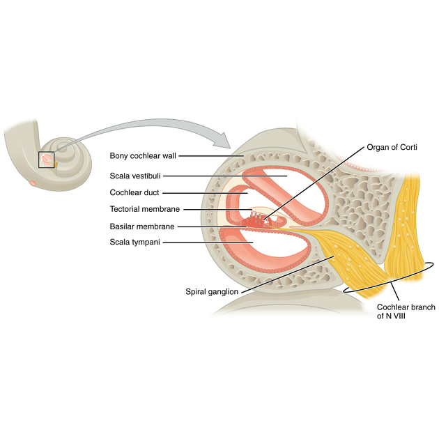
The cochlear duct (also known as the scala media) is an endolymph-filled cavity located between the scala vestibuli (upper) and the scala tympani (lower) in the cochlea which is part of the inner ear along with the vestibular apparatus 1,4. The cochlea is located in the bony labyrinth, itself found in the temporal bone 2.
On this page:
Gross anatomy
The cochlear duct is a cavity filled with endolymph and is a component of the membranous labyrinth of the ear 4. It is held in position by the lamina of the modiolus 1. The cochlear duct starts at the saccule and ends blindly at the apex of the cochlea. The cochlear duct subdivides the bony labyrinth into two perilymph chambers, namely the scala vestibuli anteriorly (opens into the vestibule) and the scala tympani posteriorly (ends at the round window) 1. It is separated from the scala vestibuli by Reissner's membrane (vestibular membrane) 5. The organ of Corti, the sensory organ for hearing, lies within the cochlear duct 2.
The cochlear duct is described as being triangular in shape and has 1,4:
- outer wall: consists of thickened periosteum, known as the spiral ligament
- roof (vestibular membrane): separates the cochlear duct from the scala vestibuli
- floor: separates the cochlear duct from the scala tympani. It also consists of the lamina modiolus and basilar membrane, which supports the organ of Corti
Arterial supply
- proper cochlear artery (main cochlear artery), a subdivision of the common cochlear artery, itself a branch of the labyrinthine artery (also known as the auditory artery or internal auditory artery) 2,4
Adequate blood supply is crucial for auditory transduction and therefore the function of the cochlea 2.
Venous drainage
Venous drainage of the cochlear duct occurs through the cochlear veins and vestibular veins 4. These merge and form the labyrinthine vein, which drains either into the sigmoid sinus or the inferior petrosal sinus 2,4.
Innervation
The cochlear nerve is one branch of the vestibulocochlear nerve (CN VIII) which innervates the cochlear duct 1,4.
Radiographic features
The cochlear duct is a structure of the membranous labyrinth that cannot be distinguished on high-resolution CT or MRI as it is too small but sits in the central area of the cochlea 3.
Related pathology
See also
References
- 1. Annerie M. A. van der Jagt, Randy K. Kalkman, Jeroen J. Briaire, Berit M. Verbist, Johan H. M. Frijns. Variations in cochlear duct shape revealed on clinical CT images with an automatic tracing method. Scientific Reports. 7 (1): 17566. doi:10.1038/s41598-017-16126-6
- 2. Shi X. Physiopathology of the cochlear microcirculation. (2011) Hearing research. 282 (1-2): 10-24. doi:10.1016/j.heares.2011.08.006 - Pubmed
- 3. Juliano AF, Ginat DT, Moonis G. Imaging review of the temporal bone: part I. Anatomy and inflammatory and neoplastic processes. (2013) Radiology. 269 (1): 17-33. doi:10.1148/radiol.13120733 - Pubmed
- 4. Richard Drake, A. Wayne Vogl, Adam W. M. Mitchell. Gray's Basic Anatomy. (2016) ISBN: 9780323508506
- 5. Hannon. Porth Pathophysiology. Chapter 55. ISBN: 9781451192896
Incoming Links
Related articles: Anatomy: Head and neck
- skeleton of the head and neck
-
cranial vault
- scalp (mnemonic)
- fontanelle
-
sutures
- calvarial
- facial
- frontozygomatic suture
- frontomaxillary suture
- frontolacrimal suture
- frontonasal suture
- temporozygomatic suture
- zygomaticomaxillary suture
- parietotemporal suture (parietomastoid suture)
- occipitotemporal suture (occipitomastoid suture)
- sphenofrontal suture
- sphenozygomatic suture
- spheno-occipital suture (not a true suture)
- lacrimomaxillary suture
- nasomaxillary suture
- internasal suture
- basal/internal
- skull landmarks
- frontal bone
- temporal bone
- parietal bone
- occipital bone
- skull base (foramina)
-
facial bones
- midline single bones
- paired bilateral bones
- cervical spine
- hyoid bone
- laryngeal cartilages
-
cranial vault
- muscles of the head and neck
- muscles of the tongue (mnemonic)
- muscles of mastication
-
facial muscles
- epicranius muscle
- circumorbital and palpebral muscles
- nasal muscles
-
buccolabial muscles
- elevators, retractors and evertors of the upper lip
- levator labii superioris alaeque nasalis muscle
- levator labii superioris muscle
- zygomaticus major muscle
- zygomaticus minor muscle
- levator anguli oris muscle
- malaris muscle
- risorius muscle
- depressors, retractors and evertors of the lower lip
- depressor labii inferioris muscle
- depressor anguli oris muscle
- mentalis muscle
- compound sphincter
-
orbicularis oris muscle
- incisivus labii superioris muscle
- incisivus labii inferioris muscle
-
orbicularis oris muscle
- muscle of mastication
- modiolus
- elevators, retractors and evertors of the upper lip
- muscles of the middle ear
- orbital muscles
- muscles of the soft palate
- pharyngeal muscles
- suprahyoid muscles
- infrahyoid muscles
- intrinsic muscles of the larynx
- muscles of the neck
- platysma muscle
- longus colli muscle
- longus capitis muscle
- scalenus anterior muscle
- scalenus medius muscle
- scalenus posterior muscle
- scalenus pleuralis muscle
- sternocleidomastoid muscle
-
suboccipital muscles
- rectus capitis posterior major muscle
- rectus capitis posterior minor muscle
- obliquus capitis superior muscle
- obliquus capitis inferior muscle
- accessory muscles of the neck
- deep cervical fascia
-
deep spaces of the neck
- anterior cervical space
- buccal space
- carotid space
- danger space
- deep cervical fascia
- infratemporal fossa
- masticator space
- parapharyngeal space
- stylomandibular tunnel
- parotid space
- pharyngeal (superficial) mucosal space
- perivertebral space
- posterior cervical space
- pterygopalatine fossa
- retropharyngeal space
- suprasternal space (of Burns)
- visceral space
- surgical triangles of the neck
- orbit
- ear
- paranasal sinuses
- upper respiratory tract
- viscera of the neck
- blood supply of the head and neck
-
arterial supply
-
common carotid artery
- carotid body
- carotid bifurcation
- subclavian artery
- variants
-
common carotid artery
- venous drainage
-
arterial supply
- innervation of the head and neck
-
cranial nerves
- olfactory nerve (CN I)
- optic nerve (CN II)
- oculomotor nerve (CN III)
- trochlear nerve (CN IV)
-
trigeminal nerve (CN V) (mnemonic)
- trigeminal ganglion
- ophthalmic division
- maxillary division
- mandibular division
- abducens nerve (CN VI)
- facial nerve (CN VII)
-
vestibulocochlear nerve (CN VIII)
- vestibular ganglion (Scarpa's ganglion)
- glossopharyngeal nerve (CN IX)
- vagus nerve (CN X)
- (spinal) accessory nerve (CN XI)
- hypoglossal nerve (CN XII)
- parasympathetic ganglia of the head and neck
- cervical sympathetic ganglia
- greater occipital nerve
- third occipital nerve
-
cervical plexus
- muscular branches
- longus capitis
- longus colli
- scalenes
- geniohyoid
- thyrohyoid
-
ansa cervicalis
- omohyoid (superior and inferior bellies separately)
- sternothyroid
- sternohyoid
- phrenic nerve
- contribution to the accessory nerve (CN XI)
- cutaneous branches
- muscular branches
- brachial plexus
- pharyngeal plexus
-
cranial nerves
- lymphatic drainage of the head and neck
- embryological development of the head and neck


 Unable to process the form. Check for errors and try again.