
Coloboma is a collective term encompassing any focal discontinuity in the structure of the eye and should not be confused with staphylomas which are due to choroidal thinning.
On this page:
Terminology
While coloboma is the collective term for any focal discontinuity in the eye's structure, many people use the term to refer to iris coloboma (which is not visible on imaging). Chorioretinal colobomas are colobomas that affect the posterior globe and are most likely to be seen on imaging.
Pathology
Embryologically, colobomas are due to the failure of closure of the choroidal fissure. The most common site of ocular coloboma is the anterior segment involving the iris, less commonly in the posterior segment involving the optic disc, choroid, or retina 4. Coloboma can also involve the eyelids, lens, and optic nerve.
Colobomas may be sporadic (typically unilateral), non-syndromic inherited or syndromic (typically bilateral).
Associations
Roberts syndrome: rare
Gorlin-Goltz syndrome: rare
Radiographic features
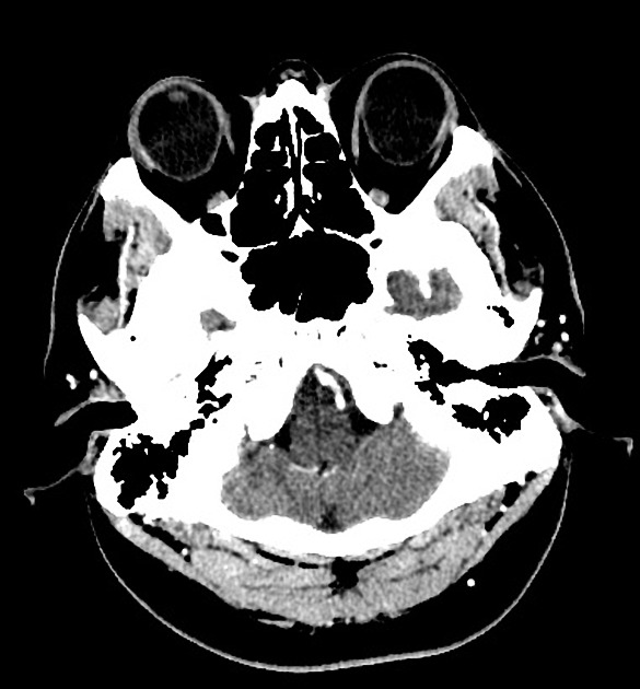
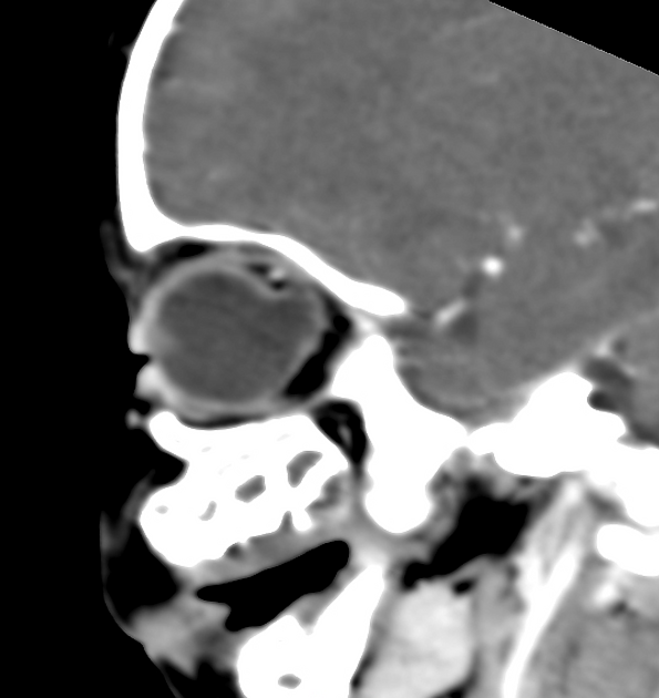
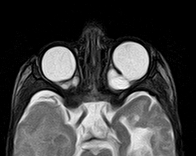
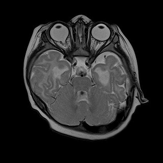
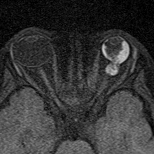
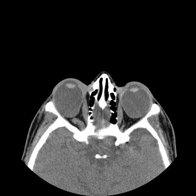
In posterior ocular coloboma, on CT or MRI, the affected globe is usually small (microphthalmia)2 with a focal posterior defect in the globe with vitreous herniation 3. A retrobulbar fluid-density cyst may be present 3.
Differential diagnosis
-
optic nerve head is funnel shaped with associated midline structural abnormalities of the brain and skull
sometimes misreported as a coloboma 7
-
microphthalmia with cyst
results from the proliferation of the embryonic retina with potential extrusion of the vitreous posteriorly into the cyst (thus microphthalmia)
the cyst may become discontinuous from the eye, be larger than the eye, and may cause proptosis 8


 Unable to process the form. Check for errors and try again.