
The contrast agent pooling sign refers to dependent contrast agent pooling in systemic veins. The original paper found a high incidence on contrast CT scans performed on emergency department patients who had a cardiac arrest within the following hour 1. Pooling is promoted by high injection rates and poor cardiac output.
On this page:
Radiographic features
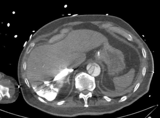
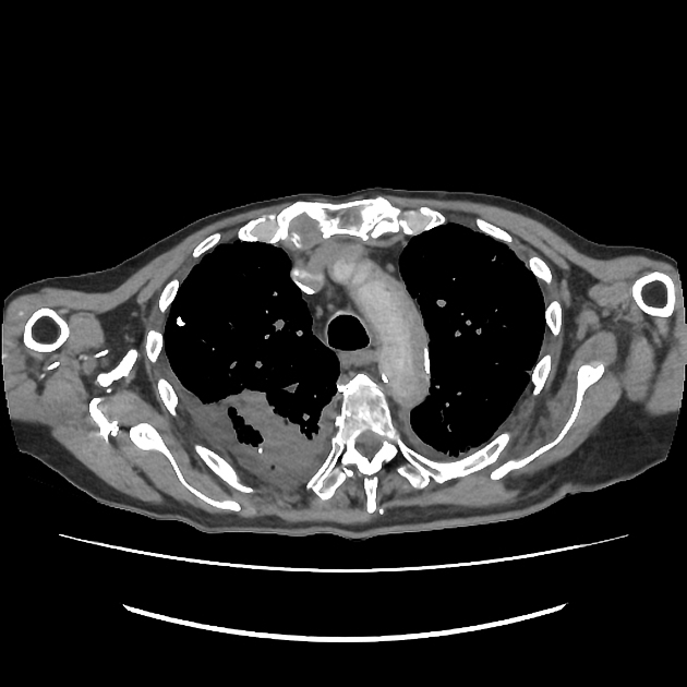
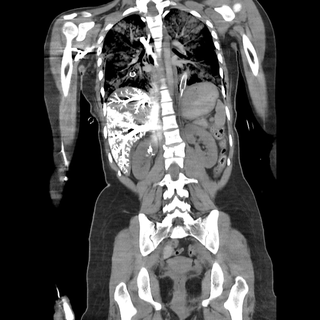
The contrast agent pooling sign is characterised by dense intravenous iodinated contrast media pooling and layering in veins such as the inferior vena cava, renal, hepatic, mesenteric, splenic, lumbar veins, or within the dependent parenchyma of the liver or kidney. Layering occasionally occurs in the superior vena cava, right atrium, right ventricle, coronary sinus, great cardiac vein, azygos vein or hemiazygos vein. Since positive CT contrast agents have a higher specific gravity than blood, they pool in the dependent part of the systemic venous system, and pooling is more extensive in patients with slow flow and cardiogenic shock 1.
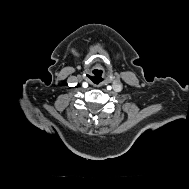
Minor contrast medium refux and layering can be normal in the internal jugular vein (retrograde flow with ipselateral upper limb contrast medium injection) but is more common and severe if the brachiocephalic vein is narrowed. Moderate pooling in the IVC and hepatic veins is quite frequent and may be of no consequence. It could be due to clinically unimportant tricuspid regurgitation, which is common and increases in incidence and severity with age 6.
Treatment and prognosis
If pathological, the underlying condition should be treated, e.g. aortic dissection, hypovolaemic shock due to trauma or bleeding, pulmonary embolism, myocardial infarction, diastolic dysfunction, constrictive pericarditis, large pleural or pericardial effusions, cerebral haemorrhage, and septic shock.
History and etymology
The dependent pooling of contrast media in the context of cardiac arrest was first described in the English language in 2002; although this article cites a German language case report from 2000, which summarises similar findings due to a cardiac arrest whilst a patient underwent CT 2. The German article does not cite any earlier reports demonstrating such a finding 3.


 Unable to process the form. Check for errors and try again.