
Dedifferentiated chondrosarcomas (DCS) are malignant high-grade chondrosarcomas with a poor prognosis. They have a bi-morphic histomorphology of conventional chondrosarcoma and a non-cartilaginous high-grade sarcoma.
On this page:
Epidemiology
Dedifferentiated chondrosarcomas can develop in 10-15% of central chondrosarcomas and a rare percentage of peripheral chondrosarcomas 1,2. They have been found in a wide age range from 15-85 years and a median age between 55-60 years if they develop from a central location 1-5. If they originate from peripheral chondrosarcoma they occur slightly earlier. The male gender is more frequently affected 1,2.
Diagnosis
The diagnosis of dedifferentiated chondrosarcoma is established histologically 1.
Diagnostic criteria
Diagnostic criteria according to the WHO classification of soft tissue and bone tumours (5th edition) 1:
bi-morphic histology of conventional chondrosarcoma and another high-grade sarcoma with a crisp interface
The presence of IDH mutation is a desirable criterion 1.
Clinical presentation
The most common symptoms of dedifferentiated chondrosarcomas are swelling/palpable mass lesion and pain. About 20% of those tumours are associated with a pathological fracture 1-3.
Pathology
Dedifferentiated chondrosarcomas are highly malignant neoplasms with a bi-morphic histomorphology consisting of chondrosarcoma of any grade juxtaposed to a high-grade non-cartilaginous sarcoma such as an undifferentiated pleomorphic sarcoma, osteosarcoma, spindle cell sarcoma, and less commonly rhabdomyosarcoma, leiomyosarcoma or angiosarcoma 1,5.
Aetiology
At the time of writing, the aetiology of dedifferentiated chondrosarcoma is unknown 1.
Location
Overall and for tumours originating from central chondrosarcoma the following locations are most frequently involved 1-3:
femur
pelvis
humerus
scapula
The most common sites of dedifferentiated chondrosarcomas arising from a peripheral chondrosarcoma 1:
pelvis
scapula
ribs
Macroscopic appearance
Grossly, dedifferentiated chondrosarcomas are characterised by varying proportions of cartilaginous and non-cartilaginous tumour components 1,2. A lobulated form usually characterises the chondroid component and a bluish-grey colour which is located more centrally the sarcomatous component appears pale-yellow to tannish-brown and is usually found in an extraosseous location or near a pathological fracture 1,2.
Microscopic appearance
Under the microscope, dedifferentiated are characterised by a bi-morphic histomorphology and an abrupt transition between cartilaginous and high-grade sarcoma components 1-3,5.
The chondroid or cartilaginous tumour components display features that range anywhere from the appearance of an enchondroma over low-grade to intermediate and high-grade chondrosarcoma 1.
The high-grade component usually displays histological features of undifferentiated pleomorphic sarcoma or osteosarcoma, less frequently those of rhabdomyosarcoma, leiomyosarcoma or angiosarcoma and in very rare occasions squamous, epithelial or adamantinoma-like features 1. The ratio between chondrosarcoma and high-grade sarcoma tumour proportion is highly variable, ranging between 2% and 98% 1.
Immunophenotype
The immunophenotype of the non-cartilaginous sarcomatous tumour component follows the differentiation line of the corresponding component with the expression of desmin or keratins in some of the tumours. Only a tiny fraction of IDH1 mutations are recognised by mutation-specific IDH1 antibodies and is therefore for limited use concerning the chondrosarcoma component, but the cartilaginous portion shows reactivity to S100 4. The dedifferentiated component of more than half of the tumours shows overexpression of p53 1,4.
Genetics
Identical TP53 and IDH mutations might be found in conventional chondrogenic and dedifferentiated sarcomatous tumour components and then indicate a common origin of both parts 1.
Radiographic features
Radiographic features of dedifferentiated chondrosarcoma resemble those of intermediate to high-grade chondrosarcoma in the majority of cases. In a minority of cases, their appearance is that of low-grade chondrosarcoma. In up to 10-15% of cases there is no radiographic evidence of a chondroid matrix or an underlying chondral tumour and some of those show features of an osteosarcoma 6,7.
A bi-morphic appearance with unmineralized tumour mass within or adjacent to mineralised chondroid tumour components has been described in up to 30% of cases on radiographs and up to 50% of cases on CT 1.
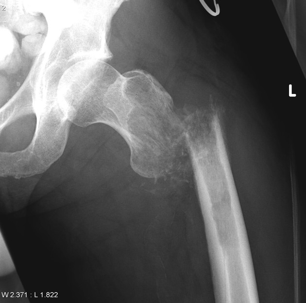
Plain radiograph/CT
Radiographic features of periosteal chondrosarcomas include the following 6,7:
osteolytic lesion with permeative and destructive areas
intralesional rings and arcs calcifications
cortical destruction
cortical thickening
periosteal reaction (most common: solid; very rarely: spiculated)
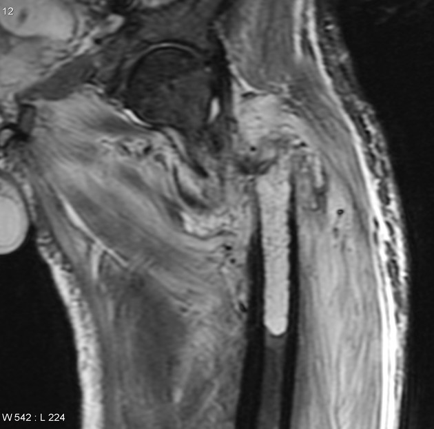
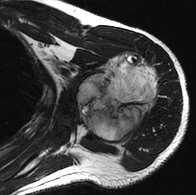
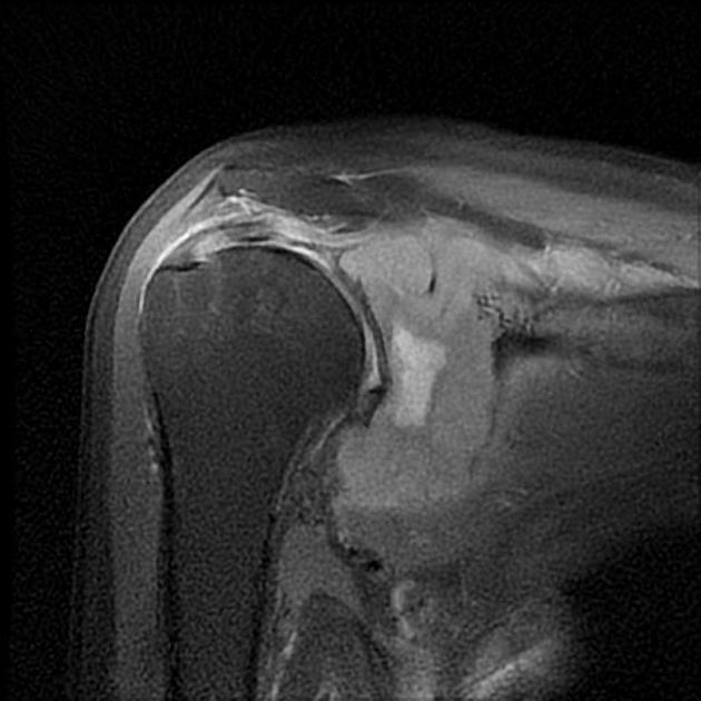
MRI
MRI might demonstrate a bi-morphic or biphasic appearance, that is the coexistence of a cartilaginous tumour with a chondroid matrix and a tumour component of an aggressive non-chondral lesion with or without osseous matrix. This includes tumour components with relatively low signal intensities on T2 and relatively uniform and/or inhomogeneous contrast enhancement patterns next to areas of a typical chondroid matrix with high signal intensity on fluid-sensitive sequences with intralesional signal voids and peripheral and/or septal enhancement patterns 6-8.
However, it should be noted, that a bi-morphic or biphasic appearance is not reliable in distinguishing dedifferentiated chondrosarcoma from a secondary peripheral chondrosarcoma or a periosteal chondrosarcoma 9.
Radiology report
The radiological report should include a description of the following 6-9:
tumour size and location (metaphysis, diaphysis)
tumour margins/surface
intralesional calcifications
cortical erosion/destruction
extension into the medullary cavity
association to the periosteum/periosteal shell
presence of a stalk
presence of metastases
Treatment and prognosis
The prognosis of dedifferentiated chondrosarcoma is poor with an overall 5-year survival rate of well below 30% 1-3,10. Treatment includes surgery with wide or radical resection 10. Radiation therapy and chemotherapy have not yet been shown to improve prognosis 1,3. Large tumour size >8cm, the presence of a pathological fracture, pelvic location, inadequate surgical margins and of course metastases are associated with poor outcomes 1.
Complications
Complications include local recurrences and metastases, especially to the lungs 1,3.
History and etymology
Dedifferentiated chondrosarcoma was first described by the American pathologists David C. Dahlin and John W. Beabout in 1971 2,11.
Differential diagnosis
Differential diagnoses of dedifferentiated chondrosarcoma include 6-9:


 Unable to process the form. Check for errors and try again.