
Doppler shift
Citation, DOI, disclosures and article data
At the time the article was created Ayush Goel had no recorded disclosures.
View Ayush Goel's current disclosuresAt the time the article was last revised Craig Hacking had the following disclosures:
- Philips Australia, Paid speaker at Philips Spectral CT events (ongoing)
These were assessed during peer review and were determined to not be relevant to the changes that were made.
View Craig Hacking's current disclosures- Doppler effect
- Doppler shifts
- Doppler effects
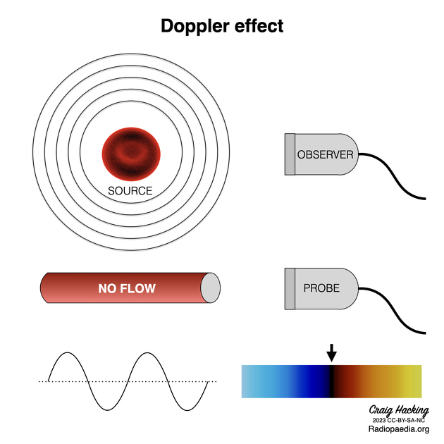
Doppler shift or Doppler effect is defined as the change in frequency of sound wave due to a reflector moving towards or away from an object, which in the case of ultrasound is the transducer.
On this page:
Images:
Terminology
When sound of a given frequency is discharged and subsequently reflected from a source that is not in motion, the frequency of the returning sound waves will equal the frequency at which they were emitted.
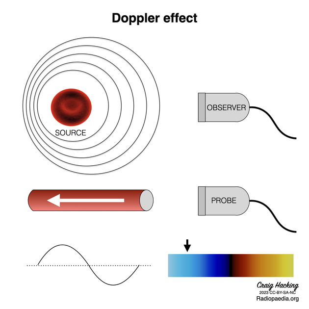
However, if the reflecting source is in motion either toward or away from the emitting source (e.g. an ultrasound transducer) the frequency of the sound waves received will be higher (positive Doppler shift) or lower (negative Doppler shift) than the frequency at which they were emitted, respectively 2.
-
positive Doppler shift
frequency of received sound waves > frequency of emitted sound waves
source reflecting sound waves is moving toward the emitting source
depicted in colour flow Doppler as red
spectral envelope (in continuous and pulsed wave Doppler) above the baseline
-
negative Doppler shift
frequency of received sound waves < frequency of emitted sound waves
source reflecting sound waves is moving away from the emitting source
depicted in colour flow Doppler as blue
spectral envelope (in continuous and pulsed wave Doppler) below the baseline
Doppler equation
F = 2fo(v/c)cos(Q)
where:
F is Doppler frequency shift
fo is transmitted frequency from ultrasound probe
v is the velocity of moving reflector
c is the velocity of sound in the medium
-
Q is the angle between ultrasound beam and axis of flow
the Greek letter theta (θ) is also used
The above doppler formula is used because the transducer is not parallel to the axis of the moving object 4.
The magnitude of the Doppler shift is affected by the angle at which the reflecting source is travelling in relation to the transmitting source. This is accounted for in the Doppler equation with the "cosine(θ)" parameter; the maximum Doppler shift occurs when the relative motion occurs at a Doppler angle of 0 degrees (the cosine of 0 = 1) and no Doppler shift will be noted when the motion of the reflecting source is perpendicular (cosine of 90 = 0) 3.
ADVERTISEMENT: Supporters see fewer/no ads
History and etymology
Named after Austrian physicist, Christian Andreas Doppler (1803-1853) 1.
See also
Quiz questions
References
- 1. Katsi V, Felekos I, Kallikazaros I. Christian Andreas Doppler: A legendary man inspired by the dazzling light of the stars. (2013) Hippokratia. 17 (2): 113-4. Pubmed
- 2. Kruskal JB, Newman PA, Sammons LG, Kane RA. Optimizing Doppler and color flow US: application to hepatic sonography. (2004) Radiographics : a review publication of the Radiological Society of North America, Inc. 24 (3): 657-75. doi:10.1148/rg.243035139 - Pubmed
- 3. Ravi Rasalingam, Majesh Makan, Julio E. Perez. The Washington Manual of Echocardiography. (2012) ISBN: 9781451113402
- 4. Keane P, Ruiz-Garcia H, Sadda S. Advanced Imaging Technologies. Retina. 2013;:133-50. doi:10.1016/b978-1-4557-0737-9.00005-9
Incoming Links
- End-diastolic velocity (Doppler ultrasound)
- Tissue Doppler imaging (echocardiography)
- Color bruit artifact
- Aliasing phenomenon (ultrasound)
- Coandă effect (physics)
- Wall filter
- Color flow Doppler (ultrasound)
- Nyquist limit
- Spectral Doppler (ultrasound)
- Color flash artifact
- History of ultrasound in medicine
- Doppler angle correction
- Power Doppler
- Peak systolic velocity (Doppler ultrasound)
Related articles: Imaging physics
- imaging physics
- imaging in practice
- imaging technology
-
x-ray physics
- ionising radiation
- interaction with matter
- x-ray spectrum
- radiation units
- effective dose
- entrance skin dose
- radiation safety
- radiation damage (biomolecular)
- radiation damage (skin injury)
- stochastic effect
- CT physics
-
MRI physics
- B0
- chemical shift
- dependence of magnetisation (proton density, field strength and temperature)
- echo time
- eddy currents
- electromagnetic induction
- Ernst angle
- flip angle
- Larmor frequency
- magnetic dipole
- magnetic field gradient
- magnetic susceptibility
- magnetism
- molecular tumbling rate effects on T1 and T2
- net magnetisation vector (NMV)
- relaxation
- repetition time
- resonance and radiofrequency (RF)
- units of magnetism
- ultrasound physics
- nuclear medicine physics
Related articles: Imaging technology
- imaging technology
- imaging physics
- imaging in practice
-
x-rays
- x-ray physics
- x-ray in practice
- x-ray production
- x-ray tube
- filters
- automatic exposure control (AEC)
- beam collimators
- grids
- air gap technique
- cassette
- intensifying screen
- x-ray film
- image intensifier
- digital radiography
- digital image
- mammography
- x-ray artifacts
- radiation units
- radiation safety
- radiation detectors
- fluoroscopy
-
computed tomography (CT)
- CT physics
- CT in practice
- CT technology
- CT image reconstruction
- CT image quality
- CT dose
-
CT contrast media
-
iodinated contrast media
- agents
- water soluble
- water insoluble
- vicarious contrast material excretion
- iodinated contrast media adverse reactions
- agents
- non-iodinated contrast media
-
iodinated contrast media
-
CT artifacts
- patient-based artifacts
- physics-based artifacts
- hardware-based artifacts
- ring artifact
- tube arcing
- out of field artifact
- air bubble artifact
- helical and multichannel artifacts
- CT safety
- history of CT
-
MRI
- MRI physics
- MRI in practice
- MRI hardware
- signal processing
-
MRI pulse sequences (basics | abbreviations | parameters)
- T1 weighted image
- T2 weighted image
- proton density weighted image
- chemical exchange saturation transfer
- CSF flow studies
- diffusion weighted imaging (DWI)
- echo-planar pulse sequences
- fat-suppressed imaging sequences
- gradient echo sequences
- inversion recovery sequences
- metal artifact reduction sequence (MARS)
-
perfusion-weighted imaging
- techniques
- derived values
- saturation recovery sequences
- spin echo sequences
- spiral pulse sequences
- susceptibility-weighted imaging (SWI)
- T1 rho
- MR angiography (and venography)
-
MR spectroscopy (MRS)
- 2-hydroxyglutarate peak: resonates at 2.25 ppm
- alanine peak: resonates at 1.48 ppm
- choline peak: resonates at 3.2 ppm
- citrate peak: resonates at 2.6 ppm
- creatine peak: resonates at 3.0 ppm
- functional MRI (fMRI)
- gamma-aminobutyric acid (GABA) peak: resonates at 2.2-2.4 ppm
- glutamine-glutamate peak: resonates at 2.2-2.4 ppm
- Hunter's angle
- lactate peak: resonates at 1.3 ppm
- lipids peak: resonates at 1.3 ppm
- myoinositol peak: resonates at 3.5 ppm
- MR fingerprinting
- N-acetylaspartate (NAA) peak: resonates at 2.0 ppm
- propylene glycol peak: resonates at 1.13 ppm
-
MRI artifacts
- MRI hardware and room shielding
- MRI software
- patient and physiologic motion
- tissue heterogeneity and foreign bodies
- Fourier transform and Nyquist sampling theorem
- MRI contrast agents
- MRI safety
-
ultrasound
- ultrasound physics
-
transducers
- linear array
- convex array
- phased array
- frame averaging (frame persistence)
- ultrasound image resolution
- imaging modes and display
- pulse-echo imaging
- real-time imaging
-
Doppler imaging
- Doppler effect
- colour Doppler
- power Doppler
- B flow
- colour box
- Doppler angle
- pulse repetition frequency and scale
- wall filter
- colour write priority
- packet size (dwell time)
- peak systolic velocity
- end-diastolic velocity
- resistive index
- pulsatility index
- Reynolds number
- panoramic imaging
- compound imaging
- harmonic imaging
- elastography
- scanning modes
- 2D ultrasound
- 3D ultrasound
- 4D ultrasound
- M-mode
-
ultrasound artifacts
- acoustic shadowing
- acoustic enhancement
- beam width artifact
- reverberation artifact
- ring down artifact
- mirror image artifact
- side lobe artifact
- speckle artifact
- speed displacement artifact
- refraction artifact
- multipath artifact
- anisotropy
- electrical interference artifact
- hardware-related artifacts
- Doppler artifacts
- aliasing
- tissue vibration
- spectral broadening
- blooming
- motion (flash) artifact
- twinkling artifact
- acoustic streaming
- biological effects of ultrasound
- history of ultrasound
-
nuclear medicine
- nuclear medicine physics
- detectors
- tissue to background ratio
-
radiopharmaceuticals
- fundamentals of radiopharmaceuticals
- radiopharmaceutical labelling
- radiopharmaceutical production
- nuclear reactor produced radionuclides
- cyclotron produced radionuclides
- radiation detection
- dosimetry
- specific agents
- carbon-11
- chromium-51
- fluorine agents
- gallium agents
- Ga-67 citrate
- Ga-68
- iodine agents
-
I-123
- I-123 iodide
- I-123 ioflupane (DaTSCAN)
- I-123 ortho-iodohippurate
- I-131
-
MIBG scans
- I-123 MIBG
- I-131 MIBG
-
I-123
- indium agents
- In-111 Octreoscan
- In-111 OncoScint
- In-111 Prostascint
- In-111 oxine labelled WBC
- krypton-81m
- nitrogen-13
- oxygen-15
- phosphorus-32
- selenium-75
-
technetium agents
- Tc-99m DMSA
- Tc-99m DTPA
- Tc-99m DTPA aerosol
- Tc-99m HMPAO
- Tc-99m HMPAO labelled WBC
- Tc-99m MAA
- Tc-99m MAG3
- Tc-99m MDP
- Tc-99m mercaptoacetyltriglycine
- Tc-99m pertechnetate
- Tc-99m labelled RBC
- Tc-99m sestamibi
- Tc-99m sulfur colloid
- Tc-99m sulfur colloid (oral)
- thallium-201 chloride
- xenon agents
- in vivo therapeutic agents
- pharmaceuticals used in nuclear medicine
-
emerging methods in medical imaging
- radiography
- phase-contrast imaging
- CT
- deep-learning reconstruction
- photon counting CT
- virtual non-contrast imaging
- ultrasound
- magnetomotive ultrasound (MMUS)
- superb microvascular imaging
- ultrafast Doppler imaging
- ultrasound localisation microscopy
- MRI
- nuclear medicine
- total body PET system
- immuno-PET
- miscellaneous
- radiography


 Unable to process the form. Check for errors and try again.