
Double inlet left ventricle (DILV) describes a congenital cardiac anomaly in which both atrioventricular valves are associated with a single ventricle demonstrating left ventricular morphology.
Epidemiology
This uncommon entity constitutes 1% of all congenital cardiac anomalies and is one of the more common variants of a univentricular heart 1.
Associations
pulmonary stenosis (may be present in 2/3's of patients 4)
Pathology
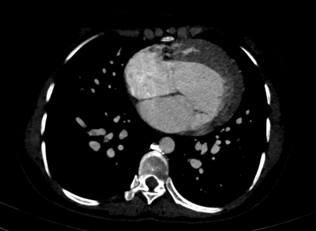
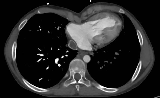
Features include a single, dominant ventricle with an elliptical shape, and smooth septal endocardium lacking associated papillary muscles, defining left ventricular morphology 3. Atrioventricular valves may lack sufficient anatomical features to determine their morphology in mitral/tricuspid valves and are commonly stenotic or hypoplastic. A rudimentary outflow chamber (right ventricle) may be identified at the cardiac base. The location of the septum may be left/anterior, defining the L-loop orientation, or right/anterior defining the less common D-loop orientation. Further classification is based upon the relation of the great arteries 1:
-
type I
normal arterial relations
-
type II
rightward/anterior aortic location
-
type III
leftward/anterior aortic location
-
type IV
leftward/posterior aortic location


 Unable to process the form. Check for errors and try again.