
Eleventh rib
Citation, DOI, disclosures and article data
Citation:
Palipana D, Hacking C, Kang O, Eleventh rib. Reference article, Radiopaedia.org (Accessed on 29 Mar 2025) https://doi.org/10.53347/rID-44310
rID:
44310
Article created:
17 Apr 2016,
Dinesh Palipana
Disclosures:
At the time the article was created Dinesh Palipana had no recorded disclosures.
View Dinesh Palipana's current disclosures
Last revised:
Disclosures:
At the time the article was last revised Craig Hacking had no recorded disclosures.
View Craig Hacking's current disclosures
Revisions:
5 times, by
3 contributors -
see full revision history and disclosures
Systems:
Sections:
The atypical 11th rib is one of two floating ribs.
Gross anatomy
Osteology
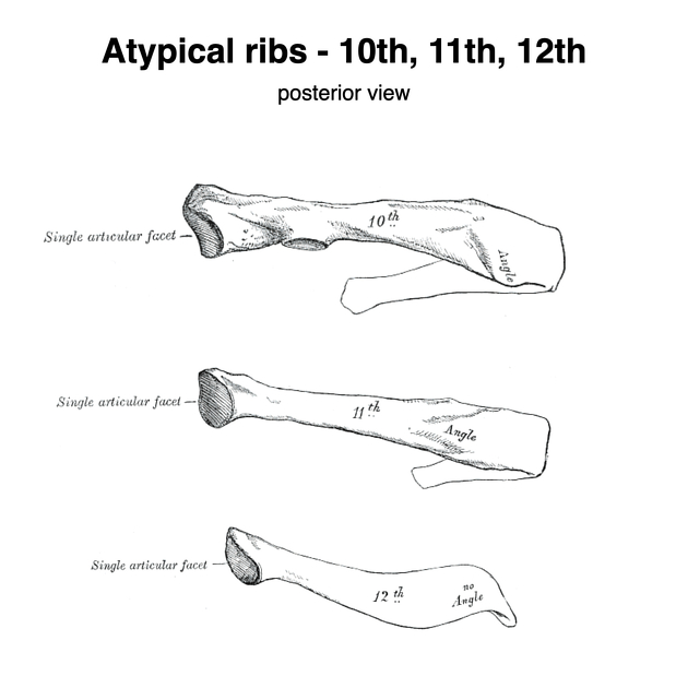
The 11th rib has a single facet on its head for articulation with the T11 vertebra. It has a short neck and no tubercle. The angle is slight. Its costal groove is shallow. The internal surface of this rib faces slightly upwards.
The pointed anterior end of the 11th rib is covered with costal cartilage. Its length is highly variable.
Related pathology
- infection, e.g. septic arthritis, osteomyelitis
- malignancy, e.g. chondrosarcoma, enchondroma, metastases
- trauma, e.g. fracture
- fracture of the eleventh rib is rare, but may be associated with haemorrhage surrounding the adrenal glands, visceral injury to the kidneys and spleen, and lumbar and thoracic vertebral injury
References
- 1. Gray's anatomy. Elsevier. ISBN:0808923714. Read it at Google Books - Find it at Amazon
- 2. Moore KL, Dalley AF. Anatomy. Lippincott Williams & Wilkins. (1999) ISBN:0683061410. Read it at Google Books - Find it at Amazon
- 3. Last's Anatomy. Churchill Livingstone. ISBN:0702033944. Read it at Google Books - Find it at Amazon
- 4. Snell RS. Clinical Anatomy by Regions. Lippincott Williams & Wilkins. ISBN:160913446X. Read it at Google Books - Find it at Amazon
- 5. Shweiki E, Klena J, Wood GC et-al. Assessing the true risk of abdominal solid organ injury in hospitalized rib fracture patients. J Trauma. 2001;50 (4): 684-8. Pubmed citation
- 6. Miller JA, Ghanekar D. Pneumothoraces secondary to blunt abdominal trauma: aids to plain film radiographic diagnosis and relationship to solid organ injury. Am Surg. 1996;62 (5): 416-20. Pubmed citation
- 7. Jabre A, Barest G, Sledge J et-al. Cord transection by guillotine effect of fractured ribs. J Trauma. 2001;50 (4): 733-4. Pubmed citation
Incoming Links
Articles:
Related articles: Anatomy: Thoracic
- thoracic skeleton
- thoracic cage
- thoracic spine
- articulations
- muscles of the thorax
- diaphragm
- intercostal space
- intercostal muscles
- variant anatomy
- spaces of the thorax
- thoracic viscera
- lower respiratory tract
-
heart
- cardiac chambers
- heart valves
- cardiac fibrous skeleton
- innervation of the heart
- development of the heart
- cardiac wall
-
pericardium
- epicardium
- epicardial fat pad
- pericardial space
- oblique pericardial sinus
- transverse pericardial sinus
-
pericardial recesses
- aortic recesses
- pulmonic recesses
- postcaval recess
- pulmonary venous recesses
- pericardial ligaments
- myocardium
- endocardium
-
pericardium
- oesophagus
- thymus
- breast
- arterial supply of the thorax
-
thoracic aorta (development)
-
ascending aorta
-
aortic root
- aortic annulus
-
coronary arteries
- coronary arterial dominance
- myocardial segments
-
left main coronary artery (LMCA)
- ramus intermedius artery (RI)
-
circumflex artery (LCx)
- obtuse marginal branches (OM1, OM2, etc))
- Kugel's artery
-
left anterior descending artery (LAD)
- diagonal branches (D1, D2, etc)
- septal perforators (S1, S2, etc)
-
right coronary artery (RCA)
- conus artery
- sinoatrial nodal artery
- acute marginal branches (AM1, AM2, etc)
- inferior interventricular artery (PDA)
- posterior left ventricular artery (PLV)
- congenital anomalies
- sinotubular junction
-
aortic root
- aortic arch
- aortic isthmus
- descending aorta
-
ascending aorta
- pulmonary trunk
-
thoracic aorta (development)
- venous drainage of the thorax
- superior vena cava (SVC)
- inferior vena cava (IVC)
-
coronary veins
-
cardiac veins which drain into the coronary sinus
- great cardiac vein
- middle cardiac vein
- small cardiac vein
- posterior vein of the left ventricle
- vein of Marshall (oblique vein of the left atrium)
- anterior cardiac veins
- venae cordis minimae (smallest cardiac veins or thebesian veins)
-
cardiac veins which drain into the coronary sinus
- pulmonary veins
- bronchial veins
- thoracoepigastric vein
- lymphatics of the thorax
- innervation of the thorax


 Unable to process the form. Check for errors and try again.