
Encapsulating peritoneal sclerosis is a rare non-malignant cause of acute or subacute small bowel obstruction. It is characterised by total or partial encasement of the small bowel within a thick fibrocollagenous membrane.
On this page:
Terminology
The condition was originally termed abdominal cocoon. The condition is popularly known as sclerosing encapsulating peritonitis, however, this is somewhat of a misnomer as inflammation is not always present 5. It has also been known as sclerosing peritonitis, encapsulating peritonitis, and peritonitis chronica fibrosa incapsulata.
Epidemiology
Encapsulating peritoneal sclerosis can occur at any age, with reports ranging from 2-day neonate to 82 years 5.
Clinical presentation
The presentation is non-specific and patients may present with vomiting, abdominal pain and/or subacute bowel obstruction 5.
Pathology
It can be idiopathic or secondary due to:
continuous ambulatory peritoneal dialysis (prevalence ~0.7%)
treatment with practolol
Various abdominal disorders such as sarcoidosis, familial Mediterranean fever, gastrointestinal malignancy, ovarian carcinoma 6, protein S deficiency, liver transplantation, fibrogenic foreign material, and luteinised ovarian thecomas are other rare causes.
Radiographic features
Plain radiograph
Abdominal radiographic appearances are nonspecific and may be normal or may show:
gas-fluid levels similar to those in patients with any other cause of small-bowel obstruction
the wall of the "cocoon" may calcify
Ultrasound
clumped bowel loops
trilaminar appearance of a hyperechoic membrane, hypoechoic bowel wall and hyperechoic bowel contents
ascites may be present 5
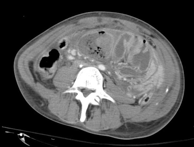
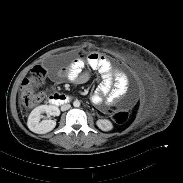
CT
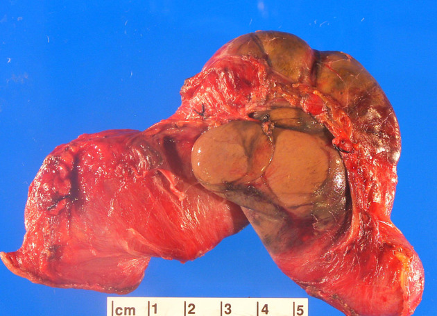
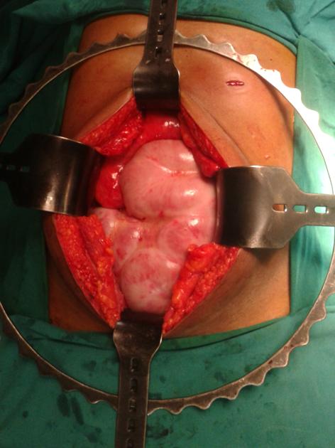
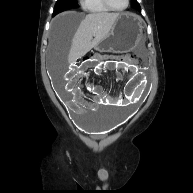
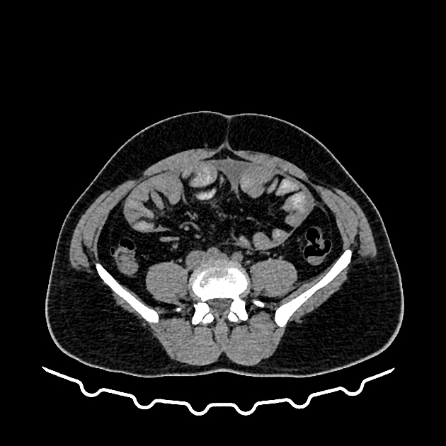
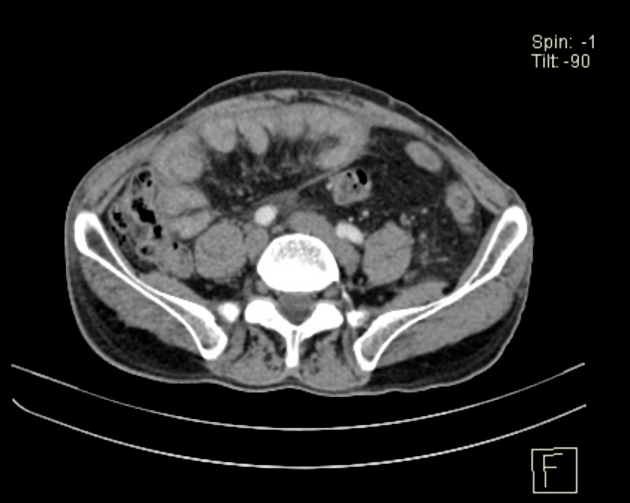
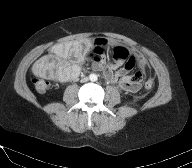
In the appropriate clinical setting, recognition of the entire dilated small bowel at the centre of the abdomen and encased within a thick fibrocollagenous membrane, as though it were in a cocoon, is diagnostic of sclerosing encapsulating peritonitis. Other imaging findings may include:
enhancing peritoneum, thickened >2 mm 5
signs of small intestinal obstruction
fixation of intestinal loops
ascites or localised fluid collections (especially interbowel)
bowel wall thickening
peritoneal or mural calcification
calcified and/or reactive adenopathy
MRI
MRI will demonstrate the same features as CT, although it may better discriminate between thickened bowel and the peritoneal membrane than CT 5.
Differential diagnosis
Encapsulating peritoneal sclerosis may be confused with congenital peritoneal encapsulation, which is characterised by a thin accessory peritoneal sac surrounding the small bowel.


 Unable to process the form. Check for errors and try again.