
Epithelioid sarcomas are malignant usually slow-growing mesenchymal tumours of unknown and multidirectional differentiation. There are classic and proximal subtypes.
On this page:
Epidemiology
Epithelioid sarcomas are rare and make up for <1% of soft tissue sarcomas 1,2. They are found in children and adults over a wide age range with the classic subtype featuring a peak incidence in adolescence and young adults and the proximal subtype showing its peak later 1. There is a male predilection. The classic subtype is apparently almost twice as common as the proximal subtype 1.
Diagnosis
Epithelioid sarcomas are diagnosed based on typical histological and molecular pathological features 1.
Diagnostic criteria
Diagnostic criteria according to the WHO classification of soft tissue and bone tumours (5th edition) 1:
- soft tissue mass (distal or proximal presentation)
- epithelioid to spindled cell morphology
- infiltrative growth
- SMARCB1/INI1 loss
- immunoreactivity for epithelial membrane antigen (EMA) and keratin
Clinical presentation
Epithelioid sarcomas usually present as single or multiple firm slow-growing mostly painless nodules, which can be associated with ulcers especially if located superficially. The proximal type is usually larger on detection 1.
Pathology
Epithelioid sarcomas show a multidirectional differentiation similar to carcinomas and feature large polygonal cells or an epithelioid to spindled cytomorphology with an infiltrative growth pattern 1.
Aetiology
The aetiology of epithelioid sarcomas is unknown. Some studies have been reporting a previous trauma which at this point is considered a probable coincidence 1.
Location
The classic or conventional subtype is usually found in the distal extremities mostly affecting the volar surfaces of the hand and fingers with proximal extremities and trunk being less common sites of occurrence 1.
The large cell or proximal type, on the other hand, is more frequently found in deep soft tissues of the pelvic girdle, the perineal genital and inguinal regions, as well as buttock and hip and can, extend along tendon sheaths and/or fasciae 1.
Subtypes
There are two accepted subtypes of epithelioid sarcomas 1:
- proximal or large cell epithelioid sarcoma
- distal or classic epithelioid sarcoma
Macroscopic appearance
Macroscopically classic epithelioid sarcomas can present as small dermal or subcutaneous nodules or multinodular lesions extending along with fasciae and nerves. They can show yellowish to brown foci due to haemorrhage and necrosis and usually feature an otherwise shimmering grey-tannish to whitish appearance. The proximal subtype is usually larger on detection 1,2.
Microscopic appearance
Histological features of epithelioid sarcomas include epithelioid cells and an infiltrative and nodular or multinodular growth pattern and differ slightly with respect to the subtype 1,2:
- classical distal subtype
- granulomatous appearance
- might show spindled tumour cells
- frequently skip lesions
- aggregates of chronic inflammatory cells
- dystrophic calcifications and metaplastic bone formation in about 1/5 of cases
- large cell or proximal subtype
- multinodular or sheet-like growth of epithelioid cells
- enlarged vesicular nuclei and prominent nucleoli
- rhabdoid features
Some epithelioid sarcomas have mixed or hybrid features of both subtypes 1.
Immunophenotype
Immunohistochemistry stains of both subtypes are usually positive for epithelial membrane antigen (EMA), vimentin and cytokeratins including CK8 and CK19 but are often negative for CK5/6. More than half of the cases show positivity of CD34 and predominantly the classic subtype shows reactivity to ERG in a fair number of cases 1,2.
Genetics
Epithelioid sarcomas are associated with mutations of the SMARCB1 or INI1 gene resulting in a loss of SMARCB1 nuclear protein expression 1.
Radiographic features
Epithelioid sarcomas have been characterised by a nodular or multinodular form and variable sometimes well-defined or irregular to ill-defined tumour margins 3,4.
US
On ultrasound, a temporally located case of epithelioid sarcoma has been described as a lobulated ill-defined hypoechoic lesion with a moderate extent of internal vascularity on colour Doppler 5.
CT
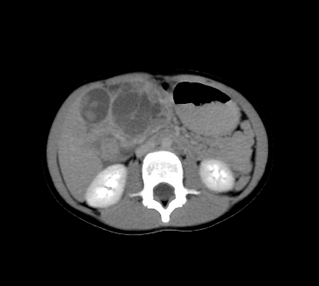
Epithelioid sarcomas have been described to show lobulated or multilobulated contours and some of the tumours have been described to show peripheral calcifications 3.
MRI
MR imaging features of epithelioid sarcomas are non-specific and feature inhomogeneous variable patterns due to variable amounts of necrosis, haemorrhage and granulomatous areas 3-6. Multiple soft tissue nodules possibly associated with ulcers should raise suspicion of this entity 4.
Signal characteristics
- T1: variable
- T2: inhomogeneous variably isointense to hyperintense compared to muscle
- T1C+ (Gd): variable inhomogeneous and heterogeneous enhancement
Nuclear medicine
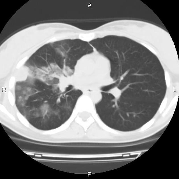
Epithelioid sarcomas have been reported to show increased uptake of FDG 5,6.
Radiology report
The radiological report should include a description of the following:
- form, location and size
- tumour margins and transition zone
- relations to the muscular fascia
- relation to adjacent neurovascular structures
- relations to bones
Treatment and prognosis
Multimodal management even of localised epithelioid sarcoma seems to have advantages in local tumour control. Local recurrence is seen in up to one-fourth of nodal involvement in one-third to half and metastatic disease in about one-third of the patients.
Overall survival rates have been reported to be in the range of 45-70% and 45-66% for 5 and 10 years respectively. Metastatic disease, nodal involvement, deep-seated tumours or proximal type, high mitotic activity, greater tumour size as well as older age of the patient are associated with a worse prognosis 1.
History and etymology
The tumour was first described by the Polish pathologist Jozef Janusz Laskowski as aponeurotic sarcoma in 1961 in the journal Nowotwory 2 and later also by the American pathologists, Bryce O. Bliss and Richard J. Reed as large cell sarcomas of tendon sheath in 1968 2,8. Eventually, the name epithelioid sarcoma was introduced by the Austrian-American pathologist Franz Michael Enzinger in 1970, who characterised them as a distinct entity 2,5-7.
Differential diagnosis
Tumours or conditions which can mimic the presentation and/or appearance of epithelioid sarcomas include 2:


 Unable to process the form. Check for errors and try again.