
Eustachian valve
Citation, DOI, disclosures and article data
At the time the article was created Matt A. Morgan had no recorded disclosures.
View Matt A. Morgan's current disclosuresAt the time the article was last revised Daniel J Bell had no financial relationships to ineligible companies to disclose.
View Daniel J Bell's current disclosures- Eustachian ridge
- Valve of the inferior vena cava
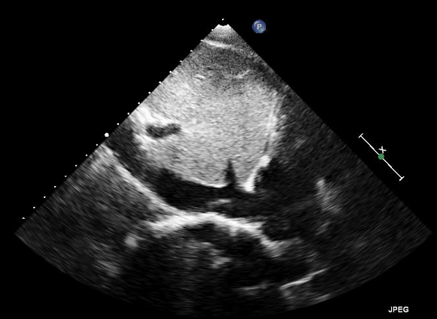
The Eustachian valve, also known as the "valve of the inferior vena cava", is a ridge of variable thickness in the inferior right atrium. It is a remnant of a fetal structure that directed incoming oxygenated blood to the foramen ovale and away from the right atrium.
Radiographic features
Incomplete regression of the Eustachian valve results in a thickened ridge at the IVC/RA junction, which can occasionally be thick enough to mimic thrombus or a right atrial mass on echocardiography, cardiac CT, or cardiac MRI.
Clinical importance
A thickened Eustachian valve may also interfere with placement of an atrial septal defect or patent foramen ovale closure device.
References
- 1. Broderick LS, Brooks GN, Kuhlman JE. Anatomic pitfalls of the heart and pericardium. Radiographics. 2005;25 (2): 441-53. doi:10.1148/rg.252045075 - Pubmed citation
- 2. Kydd AC, McNab D, Calvert PA et-al. The eustachian ridge: not an innocent bystander. JACC Cardiovasc Imaging. 2014;7 (10): 1062-3. doi:10.1016/j.jcmg.2014.02.011 - Pubmed citation
- 3. Yavuz T, Nazli C, Kinay O et-al. Giant eustachian valve with echocardiographic appearance of divided right atrium. Tex Heart Inst J. 2003;29 (4): 336-8. Free text at pubmed - Pubmed citation
Incoming Links
Related articles: Anatomy: Abdominopelvic
- skeleton of the abdomen and pelvis
- muscles of the abdomen and pelvis
- spaces of the abdomen and pelvis
- anterior abdominal wall
- posterior abdominal wall
- abdominal cavity
- pelvic cavity
- perineum
- abdominal and pelvic viscera
- gastrointestinal tract
- spleen
- hepatobiliary system
-
endocrine system
-
adrenal gland
- adrenal vessels
- chromaffin cells
- variants
- pancreas
- organs of Zuckerkandl
-
adrenal gland
-
urinary system
-
kidney
- renal pelvis
- renal sinus
- avascular plane of Brodel
-
variants
- number
- fusion
- location
- shape
- ureter
- urinary bladder
- urethra
- embryology
-
kidney
- male reproductive system
-
female reproductive system
- vulva
- vagina
- uterus
- adnexa
- Fallopian tubes
- ovaries
- broad ligament (mnemonic)
- variant anatomy
- embryology
- blood supply of the abdomen and pelvis
- arteries
-
abdominal aorta
- inferior phrenic artery
- coeliac artery
- superior mesenteric artery
- middle suprarenal artery
- renal artery (variant anatomy)
- gonadal artery (ovarian artery | testicular artery)
- inferior mesenteric artery
- lumbar arteries
- median sacral artery
-
common iliac artery
- external iliac artery
-
internal iliac artery (mnemonic)
- anterior division
- umbilical artery
- superior vesical artery
- obturator artery
- vaginal artery
- inferior vesical artery
- uterine artery
- middle rectal artery
-
internal pudendal artery
- inferior rectal artery
-
perineal artery
- posterior scrotal artery
- transverse perineal artery
- artery to the bulb
- deep artery of the penis/clitoris
- dorsal artery of the penis/clitoris
- inferior gluteal artery
- posterior division (mnemonic)
- variant anatomy
- anterior division
-
abdominal aorta
- portal venous system
- veins
- anastomoses
- arterioarterial anastomoses
- portal-systemic venous collateral pathways
- watershed areas
- arteries
- lymphatics
- innervation of the abdomen and pelvis
- thoracic splanchnic nerves
- lumbar plexus
-
sacral plexus
- lumbosacral trunk
- sciatic nerve
- superior gluteal nerve
- inferior gluteal nerve
- nerve to piriformis
- perforating cutaneous nerve
- posterior femoral cutaneous nerve
- parasympathetic pelvic splanchnic nerves
- pudendal nerve
- nerve to quadratus femoris and inferior gemellus muscles
- nerve to internal obturator and superior gemellus muscles
- autonomic ganglia and plexuses


 Unable to process the form. Check for errors and try again.