
External carotid artery
Citation, DOI, disclosures and article data
At the time the article was created Frank Gaillard had no recorded disclosures.
View Frank Gaillard's current disclosuresAt the time the article was last revised Joshua Yap had no financial relationships to ineligible companies to disclose.
View Joshua Yap's current disclosures- External carotid artery (ECA)
- ECA
- External carotid arteries
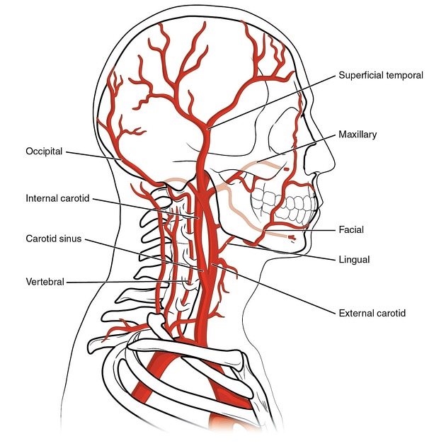
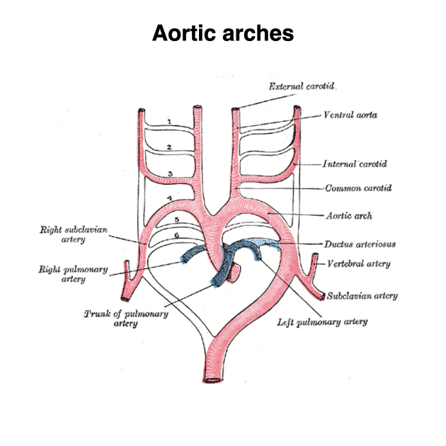
The external carotid artery (ECA) is one of the two terminal branches of the common carotid artery that has many branches that supplies the structures of the neck, face and head. The other terminal branch is the internal carotid (ICA), which is somewhat larger than the ECA, which supplies the intracranial structures.
On this page:
Summary
- origin: bifurcation of the common carotid artery
- course: under the submandibular gland and into the parotid gland
- branches:
- supply: neck, face and base of skull
- termination: division into (internal) maxillary artery and superficial temporal artery
Gross anatomy
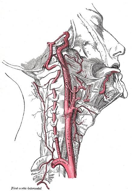
Origin and course
The ECA begins at the level of the upper border of the thyroid cartilage (at the level of the fourth cervical vertebra). It takes a slightly curved course upwards and anteriorly before inclining backwards to the space behind the neck of the mandible. Along its course, it rapidly diminishes in size and as it does so, gives off various branches (see below). As it enters the parotid gland, it gives rise to its terminal branches, the superficial temporal and maxillary arteries.
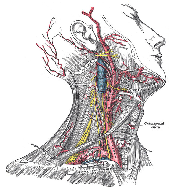
Branches
The branches of the external carotid artery can be subdivided into groups:
- arising from the carotid triangle
- posterior auricular artery
- terminal branches
Memorable mnemonics for these branches include:
- Some Anatomists Like Freaking Out Poor Medical Students
- Some American Ladies Found Our Pyramids Most Satisfactory
Relations
- anteriorly (i.e. ECA is crossed by these structures)
- upper root of ansa cervicalis
- hypoglossal nerve (CN XII)
- posterior belly of digastric muscle
- stylohyoid muscle and ligament
- common facial vein
- facial nerve (CN VII) (within the parotid gland)
- retromandibular vein (within the parotid gland)
- passing between ECA and ICA
- pharyngeal branch of vagus nerve (CN X)
- glossopharyngeal nerve (CN IX)
- stylopharyngeus muscle
- styloglossus muscle
- posteriorly (i.e. ECA lies on these structures)
- pharyngeal wall
- superior laryngeal branch of vagus nerve (CN X)
- deep lobe of the parotid gland
Variant anatomy
- variations in origin arise from the anomalous bifurcation of the common carotid artery - bifurcations may commonly be seen at the level of the cricoid cartilage (C5 level) or at the hyoid bone (C2 level)
- variant branching patterns
- linguofacial trunk (incidence ~20%): common origin lingual and facial arteries
- thyrolingual trunk (incidence ~2.5%): common origin superior thyroid and lingual arteries
- thyrolinguofacial trunk (incidence ~2.5%): common origin superior thyroid, lingual and facial arteries
- common occipito-auricular trunk (incidence ~12.5%): common origin occipital and posterior auricular arteries
References
- 1. Standring S (editor). Gray's Anatomy (39th edition). Churchill Livingstone. (2011) ISBN:0443066841. Read it at Google Books - Find it at Amazon
- 2. Schünke M, Schulte E, Ph.D. LM et-al. Atlas of anatomy, Head and neuroanatomy. George Thieme Verlag. (2007) ISBN:3131421215. Read it at Google Books - Find it at Amazon
- 3. Whitaker RH, Borley NR. Instant anatomy. Wiley-Blackwell. (2000) ISBN:0632054034. Read it at Google Books - Find it at Amazon
- 4. Thwin SS, Soe MM, Myint M et-al. Variations of the origin and branches of the external carotid artery in a human cadaver. Singapore Med J. 2010;51 (2): e40-2. Pubmed citation
Incoming Links
- Peak systolic velocity (Doppler ultrasound)
- Inferior pharyngeal constrictor muscle
- Mandible
- Antihelix (ear)
- Moyamoya disease
- Facial muscles
- Pharyngeal muscles
- Juvenile nasopharyngeal angiofibroma
- Thyroid gland
- Maxillary artery
- Giant cell arteritis
- Occipital artery
- Retromandibular vein
- Stylopharyngeus muscle
- Dural arteriovenous fistula
- Superior thyroid artery
- Notch sign (disambiguation)
- End-diastolic velocity (Doppler ultrasound)
- Caroticocavernous fistula
- Vagal paraganglioma
- Complete occlusion of left common carotid artery
- Cirsoid aneurysm
- Scalp arteriovenous malformation
- Vagal schwannoma
- Post-traumatic cirsoid aneurysm
- Anomalous separate origins of the right internal and external carotid arteries from the innominate artery
- Temporal subcutaneous high-flow arteriovenous malformation
- Carotid artery development (Gray's illustration)
- External carotid artery (Gray's illustration)
- External carotid artery main branches
- Giant scalp arteriovenous malformation
- Aortic dissection - Stanford type A
- Vagal paraganglioma
- Infantile haemangioma - parotid
- Ascending pharyngeal artery arising from internal carotid artery
- Bilateral carotid body tumours
- Vertebral artery variant: external carotid artery origin
- Normal carotid Doppler ultrasound
- Branches of the external carotid artery (Gray's anatomy illustration)
- Severe extracranial carotid artery disease
Related articles: Anatomy: Head and neck
- skeleton of the head and neck
-
cranial vault
- scalp (mnemonic)
- fontanelle
-
sutures
- calvarial
- facial
- frontozygomatic suture
- frontomaxillary suture
- frontolacrimal suture
- frontonasal suture
- temporozygomatic suture
- zygomaticomaxillary suture
- parietotemporal suture (parietomastoid suture)
- occipitotemporal suture (occipitomastoid suture)
- sphenofrontal suture
- sphenozygomatic suture
- spheno-occipital suture (not a true suture)
- lacrimomaxillary suture
- nasomaxillary suture
- internasal suture
- basal/internal
- skull landmarks
- frontal bone
- temporal bone
- parietal bone
- occipital bone
- skull base (foramina)
-
facial bones
- midline single bones
- paired bilateral bones
- cervical spine
- hyoid bone
- laryngeal cartilages
-
cranial vault
- muscles of the head and neck
- muscles of the tongue (mnemonic)
- muscles of mastication
-
facial muscles
- epicranius muscle
- circumorbital and palpebral muscles
- nasal muscles
-
buccolabial muscles
- elevators, retractors and evertors of the upper lip
- levator labii superioris alaeque nasalis muscle
- levator labii superioris muscle
- zygomaticus major muscle
- zygomaticus minor muscle
- levator anguli oris muscle
- malaris muscle
- risorius muscle
- depressors, retractors and evertors of the lower lip
- depressor labii inferioris muscle
- depressor anguli oris muscle
- mentalis muscle
- compound sphincter
-
orbicularis oris muscle
- incisivus labii superioris muscle
- incisivus labii inferioris muscle
-
orbicularis oris muscle
- muscle of mastication
- modiolus
- elevators, retractors and evertors of the upper lip
- muscles of the middle ear
- orbital muscles
- muscles of the soft palate
- pharyngeal muscles
- suprahyoid muscles
- infrahyoid muscles
- intrinsic muscles of the larynx
- muscles of the neck
- platysma muscle
- longus colli muscle
- longus capitis muscle
- scalenus anterior muscle
- scalenus medius muscle
- scalenus posterior muscle
- scalenus pleuralis muscle
- sternocleidomastoid muscle
-
suboccipital muscles
- rectus capitis posterior major muscle
- rectus capitis posterior minor muscle
- obliquus capitis superior muscle
- obliquus capitis inferior muscle
- accessory muscles of the neck
- deep cervical fascia
-
deep spaces of the neck
- anterior cervical space
- buccal space
- carotid space
- danger space
- deep cervical fascia
- infratemporal fossa
- masticator space
- parapharyngeal space
- stylomandibular tunnel
- parotid space
- pharyngeal (superficial) mucosal space
- perivertebral space
- posterior cervical space
- pterygopalatine fossa
- retropharyngeal space
- suprasternal space (of Burns)
- visceral space
- surgical triangles of the neck
- orbit
- ear
- paranasal sinuses
- upper respiratory tract
- viscera of the neck
- blood supply of the head and neck
-
arterial supply
-
common carotid artery
- carotid body
- carotid bifurcation
- subclavian artery
- variants
-
common carotid artery
- venous drainage
-
arterial supply
- innervation of the head and neck
-
cranial nerves
- olfactory nerve (CN I)
- optic nerve (CN II)
- oculomotor nerve (CN III)
- trochlear nerve (CN IV)
-
trigeminal nerve (CN V) (mnemonic)
- trigeminal ganglion
- ophthalmic division
- maxillary division
- mandibular division
- abducens nerve (CN VI)
- facial nerve (CN VII)
-
vestibulocochlear nerve (CN VIII)
- vestibular ganglion (Scarpa's ganglion)
- glossopharyngeal nerve (CN IX)
- vagus nerve (CN X)
- (spinal) accessory nerve (CN XI)
- hypoglossal nerve (CN XII)
- parasympathetic ganglia of the head and neck
- cervical sympathetic ganglia
- greater occipital nerve
- third occipital nerve
-
cervical plexus
- muscular branches
- longus capitis
- longus colli
- scalenes
- geniohyoid
- thyrohyoid
-
ansa cervicalis
- omohyoid (superior and inferior bellies separately)
- sternothyroid
- sternohyoid
- phrenic nerve
- contribution to the accessory nerve (CN XI)
- cutaneous branches
- muscular branches
- brachial plexus
- pharyngeal plexus
-
cranial nerves
- lymphatic drainage of the head and neck
- embryological development of the head and neck


 Unable to process the form. Check for errors and try again.