
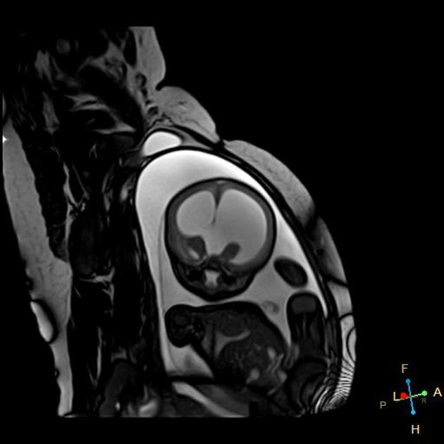
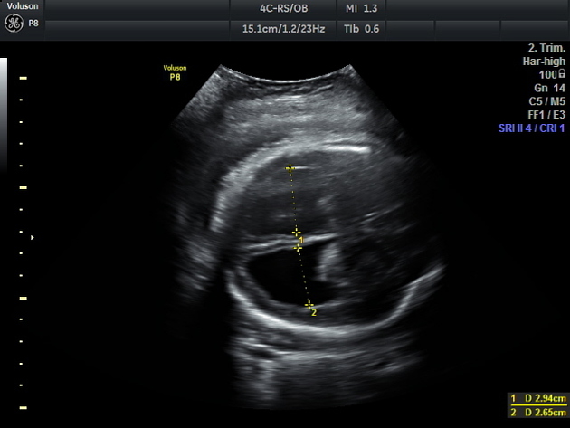
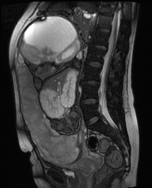
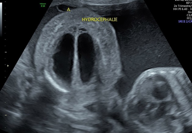
Fetal hydrocephalus often refers to an extension of fetal ventriculomegaly where the ventricular dilatation is more severe. It is usually defined when the fetal lateral ventricular diameter is >15 mm 1.
On this page:
Epidemiology
The estimated incidence is 0.5-3 cases per 1000 live births. There may be a very slight increased female predilection 11.
Pathology
Fetal hydrocephalus can be either obstructive or non-obstructive and each can arise from a number of aetiologies. In a small proportion of cases, it carries a familial X-linked inheritance (congenital X-linked hydrocephalus).
Causes
Associations
The vast majority of conditions are associated with other intracranial and cranial anomalies 6. The list includes:
-
central nervous system anomalies: (more common 7 and reported in >80% of cases)
aqueductal stenosis: one of the most common causative associations
-
non-central nervous system anomalies
-
craniofacial
facial bone anomalies
gastrointestinal anomalies
-
genitourinary anomalies
-
skeletal anomalies
-
-
syndromes
-
chromosomal anomalies: may be present in ~20% of cases 11
See also: congenital syndromes associated with enlarged ventricles.
Radiographic features
Ultrasound
Will demonstrate enlarged ventricles with variable degrees of parenchymal thinning. The choroid may be seen floating within the ventricle giving a dangling choroid sign. Often a separation of more than 3 mm between the choroid plexus and the margin of the ventricle is considered abnormal ref. In some cases, there may also be evidence of macrocephaly ref.
Treatment and prognosis
The overall prognosis will depend on the underlying cause and associated anomalies. Some cases can slowly progress during the fetal period. Antenatal shunting has been considered in a small proportion of selected cases 2.


 Unable to process the form. Check for errors and try again.