
Fetal intracranial haemorrhage may occur either within the cerebral ventricles, subdural space or infratentorial fossa.
On this page:
Pathology
Haemorrhages can occur in a number of situations:
mechanical trauma, e.g. maternal abdominal blunt or birth trauma
severe fetal hypoxia
background fetal thrombocytopenia, e.g. congenital factor X and factor V deficiencies
-
maternal thrombocytopenia
alloimmune and idiopathic thrombocytopenia
specific medications, e.g. warfarin, illicit drug (cocaine) use
haemorrhage into a fetal intracranial tumour
Radiographic feautures
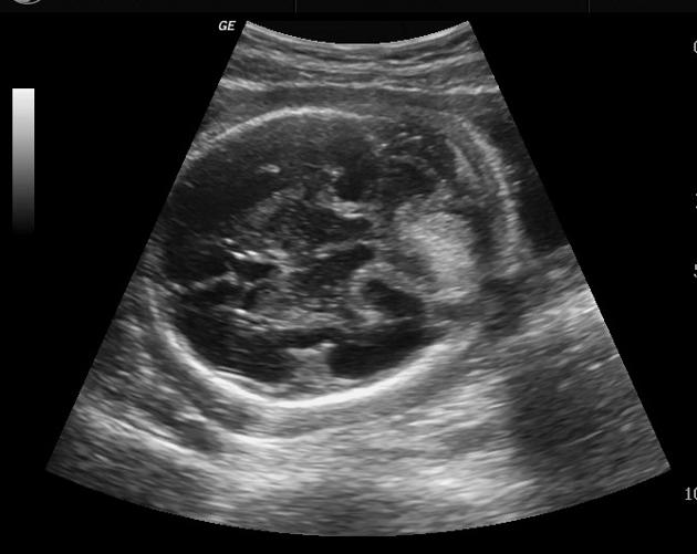
Ultrasound
The sonographic appearance of fetal intracranial haemorrhage is extremely variable, depending on its location and age of the haemorrhage. A massive intraparenchymal haemorrhage can sometimes be seen as an irregular hyperechoic mass. As the haemorrhage matures, porencephalic cyst formation or fetal intracranial calcification may be seen.
Treatment and prognosis
The outcome is usually poor particularly with parenchymal and subdural haemorrhage, whereas it is better in the subgroup with intraventricular haemorrhage.


 Unable to process the form. Check for errors and try again.