
Filters
Citation, DOI, disclosures and article data
At the time the article was created Ayush Goel had no recorded disclosures.
View Ayush Goel's current disclosuresAt the time the article was last revised Mateusz Wilczek had no financial relationships to ineligible companies to disclose.
View Mateusz Wilczek's current disclosures- Filter
- Filtration
- Hardening
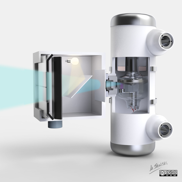
Filters are metal sheets placed in the x-ray beam between the window and the patient that are used to attenuate the low-energy (soft) x-ray photons from the spectrum. Filtering is the removal of these low energy x-rays from the beam spectrum which would otherwise not contribute to image quality but would add to patient dose and scatter. If unfiltered these low-energy x-ray photons are generally absorbed by superficial structures of the body and contribute to the entrance surface dose (ESD). As they are absorbed by the superficial structures they contribute minimally to image formation. Using a filter reduces the ESD and to a lesser extent effective dose for the patient 1.
The strength of filtration is commonly expressed in mm of aluminium half-value layer (HVL) in the filtered beam (this is a different measure than the thickness of the filter itself, even if it is made of aluminium) 7.
There are two types of filtration 2:
inherent filtration from components in the x-ray tube, i.e. window, housing, cooling oil, anode target itself (resulting in around 0.5-1.0 mm Al HVL)
added filtration from interchangeable metal sheets (Al, Cu, etc.)
Total filtration is the combined effect of inherent and added filtration, with US guidelines stating a minimum total filtration HVL of 2.5 mm of aluminium is required for x-ray tubes operating above 70 kVp 3.
The added filtration component is customised (filter thickness, type of metal) for individual examinations and procedures (e.g fluoroscopy) and takes advantage of specific metals filtration characteristics (e.g. absorption edges) to improve image quality and contrast 4.
Aluminium and copper are the most commonly used filter materials for general purpose x-ray systems. Molybdenum and rhodium filters are commonly used in mammography, where low energy beams are required 7.
Filtration reduces x-ray intensity, but not the maximum energy of the x-ray beam spectrum. The change in the shape of the beam spectrum with filtration is referred to as beam hardening. This is due to the loss of lower energy photons from a polychromatic beam. The average x-ray energy is increased and becomes more penetrating 6.
References
- 1. Brosi P, Stuessi A, Verdun F, Vock P, Wolf R. Copper Filtration in Pediatric Digital X-Ray Imaging: Its Impact on Image Quality and Dose. Radiol Phys Technol. 2011;4(2):148-55. doi:10.1007/s12194-011-0115-4
- 2. Leonie Munro, World Health Organization. Basics of Radiation Protection How to Achieve Alara. (2004) ISBN: 9789241591782
- 3. Terri L. Fauber. Radiographic Imaging and Exposure - E-Book. (2016) ISBN: 9780323443913
- 4. Peter Hertrich. Practical Radiography: Principles and Applications. (2005) ISBN: 9783895782107
- 5. Dowdey, James E., Murry, Robert C., Christensen, Edward E., 1929-. Christensen's Physics of Diagnostic Radiology. (1990) ISBN: 0812113101
- 6. Walter Huda, Richard M. Slone. Review of Radiological Physics. (2003) ISBN: 9780781736756
- 7. Robert Fosbinder, Denise Orth. Essentials of Radiologic Science. (2011) ISBN: 9780781775540 - Google Books
Incoming Links
Related articles: Imaging technology
- imaging technology
- imaging physics
- imaging in practice
-
x-rays
- x-ray physics
- x-ray in practice
- x-ray production
- x-ray tube
- filters
- automatic exposure control (AEC)
- beam collimators
- grids
- air gap technique
- cassette
- intensifying screen
- x-ray film
- image intensifier
- digital radiography
- digital image
- mammography
- x-ray artifacts
- radiation units
- radiation safety
- radiation detectors
- fluoroscopy
-
computed tomography (CT)
- CT physics
- CT in practice
- CT technology
- CT image reconstruction
- CT image quality
- CT dose
-
CT contrast media
-
iodinated contrast media
- agents
- water soluble
- water insoluble
- vicarious contrast material excretion
- iodinated contrast media adverse reactions
- agents
- non-iodinated contrast media
-
iodinated contrast media
-
CT artifacts
- patient-based artifacts
- physics-based artifacts
- hardware-based artifacts
- ring artifact
- tube arcing
- out of field artifact
- air bubble artifact
- helical and multichannel artifacts
- CT safety
- history of CT
-
MRI
- MRI physics
- MRI in practice
- MRI hardware
- signal processing
-
MRI pulse sequences (basics | abbreviations | parameters)
- T1 weighted image
- T2 weighted image
- proton density weighted image
- chemical exchange saturation transfer
- CSF flow studies
- diffusion weighted imaging (DWI)
- echo-planar pulse sequences
- fat-suppressed imaging sequences
- gradient echo sequences
- inversion recovery sequences
- metal artifact reduction sequence (MARS)
-
perfusion-weighted imaging
- techniques
- derived values
- saturation recovery sequences
- spin echo sequences
- spiral pulse sequences
- susceptibility-weighted imaging (SWI)
- T1 rho
- MR angiography (and venography)
-
MR spectroscopy (MRS)
- 2-hydroxyglutarate peak: resonates at 2.25 ppm
- alanine peak: resonates at 1.48 ppm
- choline peak: resonates at 3.2 ppm
- citrate peak: resonates at 2.6 ppm
- creatine peak: resonates at 3.0 ppm
- functional MRI (fMRI)
- gamma-aminobutyric acid (GABA) peak: resonates at 2.2-2.4 ppm
- glutamine-glutamate peak: resonates at 2.2-2.4 ppm
- Hunter's angle
- lactate peak: resonates at 1.3 ppm
- lipids peak: resonates at 1.3 ppm
- myoinositol peak: resonates at 3.5 ppm
- MR fingerprinting
- N-acetylaspartate (NAA) peak: resonates at 2.0 ppm
- propylene glycol peak: resonates at 1.13 ppm
-
MRI artifacts
- MRI hardware and room shielding
- MRI software
- patient and physiologic motion
- tissue heterogeneity and foreign bodies
- Fourier transform and Nyquist sampling theorem
- MRI contrast agents
- MRI safety
-
ultrasound
- ultrasound physics
-
transducers
- linear array
- convex array
- phased array
- frame averaging (frame persistence)
- ultrasound image resolution
- imaging modes and display
- pulse-echo imaging
- real-time imaging
-
Doppler imaging
- Doppler effect
- colour Doppler
- power Doppler
- B flow
- colour box
- Doppler angle
- pulse repetition frequency and scale
- wall filter
- colour write priority
- packet size (dwell time)
- peak systolic velocity
- end-diastolic velocity
- resistive index
- pulsatility index
- Reynolds number
- panoramic imaging
- compound imaging
- harmonic imaging
- elastography
- scanning modes
- 2D ultrasound
- 3D ultrasound
- 4D ultrasound
- M-mode
-
ultrasound artifacts
- acoustic shadowing
- acoustic enhancement
- beam width artifact
- reverberation artifact
- ring down artifact
- mirror image artifact
- side lobe artifact
- speckle artifact
- speed displacement artifact
- refraction artifact
- multipath artifact
- anisotropy
- electrical interference artifact
- hardware-related artifacts
- Doppler artifacts
- aliasing
- tissue vibration
- spectral broadening
- blooming
- motion (flash) artifact
- twinkling artifact
- acoustic streaming
- biological effects of ultrasound
- history of ultrasound
-
nuclear medicine
- nuclear medicine physics
- detectors
- tissue to background ratio
-
radiopharmaceuticals
- fundamentals of radiopharmaceuticals
- radiopharmaceutical labelling
- radiopharmaceutical production
- nuclear reactor produced radionuclides
- cyclotron produced radionuclides
- radiation detection
- dosimetry
- specific agents
- carbon-11
- chromium-51
- fluorine agents
- gallium agents
- Ga-67 citrate
- Ga-68
- iodine agents
-
I-123
- I-123 iodide
- I-123 ioflupane (DaTSCAN)
- I-123 ortho-iodohippurate
- I-131
-
MIBG scans
- I-123 MIBG
- I-131 MIBG
-
I-123
- indium agents
- In-111 Octreoscan
- In-111 OncoScint
- In-111 Prostascint
- In-111 oxine labelled WBC
- krypton-81m
- nitrogen-13
- oxygen-15
- phosphorus-32
- selenium-75
-
technetium agents
- Tc-99m DMSA
- Tc-99m DTPA
- Tc-99m DTPA aerosol
- Tc-99m HMPAO
- Tc-99m HMPAO labelled WBC
- Tc-99m MAA
- Tc-99m MAG3
- Tc-99m MDP
- Tc-99m mercaptoacetyltriglycine
- Tc-99m pertechnetate
- Tc-99m labelled RBC
- Tc-99m sestamibi
- Tc-99m sulfur colloid
- Tc-99m sulfur colloid (oral)
- thallium-201 chloride
- xenon agents
- in vivo therapeutic agents
- pharmaceuticals used in nuclear medicine
-
emerging methods in medical imaging
- radiography
- phase-contrast imaging
- CT
- deep-learning reconstruction
- photon counting CT
- virtual non-contrast imaging
- ultrasound
- magnetomotive ultrasound (MMUS)
- superb microvascular imaging
- ultrafast Doppler imaging
- ultrasound localisation microscopy
- MRI
- nuclear medicine
- total body PET system
- immuno-PET
- miscellaneous
- radiography


 Unable to process the form. Check for errors and try again.