
Flexor digitorum superficialis muscle
Citation, DOI, disclosures and article data
At the time the article was created Henry Knipe had no recorded disclosures.
View Henry Knipe's current disclosuresAt the time the article was last revised Mostafa Elfeky had no financial relationships to ineligible companies to disclose.
View Mostafa Elfeky's current disclosures- Flexor digitorum superficialis (FDS) muscles
- Flexor digitorum sublimis (FDS) muscle
- Flexor digitorum sublimis (FDS) muscles
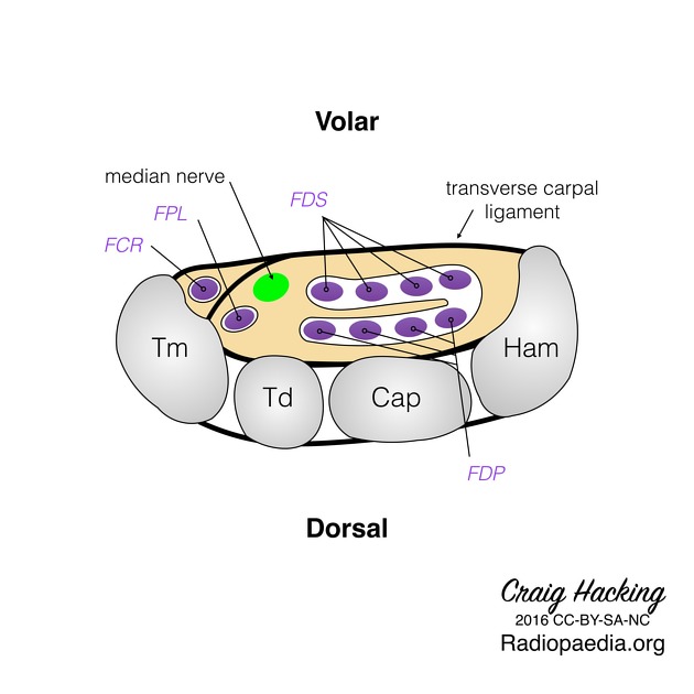
Flexor digitorum superficialis (FDS) muscle, also known as flexor digitorum sublimis muscle, is a muscle in the second (intermediate) layer of the anterior compartment of the forearm. It splits into four tendons, passes through the carpal tunnel under the flexor retinaculum. At the level of the head of the metacarpal, the flexor digitorum superficialis tendons split and wrap around the flexor digitorum profundus muscle creating an aperture for the flexor digitorum profundus to travel through. This crossing of the tendons can be referred to as the Camper chiasm. The flexor digitorum superficialis tendons rejoin deep to the flexor digitorum profundus and insert at the volar side of the proximal portion of the middle phalanx of the 2nd- 5th digits. It is one of the extrinsic muscles of the hand.
Summary
-
origin
- humeroulnar head: medial epicondyle of the humerus and coronoid process of the ulna
- radial head: diaphysis of the radius
- insertion: via four tendons into the volar aspect of the base of the middle phalanx of digits 2-5 (index, middle, ring and little fingers)
- innervation: median nerve (C7-T1)
- action: flexion of the proximal interphalangeal joints
References
- 1. Michael Schünke, Lawrence M. Ross, Erik Schulte et al. General Anatomy and Musculoskeletal System. (2010) ISBN: 9781604062922 - Google Books
Incoming Links
- Wrist
- Carpal tunnel syndrome
- Ulnar artery
- Anterior compartment of the forearm
- Scaphoid
- Trigger finger
- Kirner deformity
- Carpal tunnel
- Medical abbreviations and acronyms (F)
- Gantzer muscle
- MRI of the wrist (an approach)
- Pronator teres syndrome
- Extrinsic muscles of the hand
- Radius
- Elbow
- Zone classification of flexor tendon injury
- Median nerve
- Common flexor origin of the elbow
- Ulna
- Anterior interosseous nerve syndrome
- Flexor tendons tenosynovitis - wrist
- Flexor pulleys of the finger - anatomy
- Flexor digitorum profundus tendon tear
- Flexor digitorum profundus tendon tear (little finger)
- Flexor digitorum profundus tendon tear (little finger)
- Flexor tendons laceration - hand
- Flexor tendons laceration - hand
- Multi-ligamentous injury of the elbow with instability
- Middle finger flexor tendons partial thickness tear
- Jersey fingers
- Flexor tendons tenosynovitis - wrist
- Flexor tenosynovitis - hand
Related articles: Anatomy: Upper limb
-
skeleton of the upper limb
- clavicle
- scapula
- humerus
- radius
- ulna
- hand
- accessory ossicles of the upper limb
- accessory ossicles of the shoulder
- accessory ossicles of the elbow
-
accessory ossicles of the wrist (mnemonic)
- os centrale carpi
- os epilunate
- os epitriquetrum
- os styloideum
- os hamuli proprium
- lunula
- os triangulare
- trapezium secondarium
- os paratrapezium
- os radiostyloideum (persistent radial styloid)
- joints of the upper limb
-
pectoral girdle
-
shoulder joint
- articulations
- associated structures
- joint capsule
- bursae
- ligaments
- movements
- scapulothoracic joint
-
glenohumeral joint
- arm flexion
- arm extension
- arm abduction
- arm adduction
- arm internal rotation (medial rotation)
- arm external rotation (lateral rotation)
- circumduction
- arterial supply - scapular anastomosis
- ossification centres
-
shoulder joint
-
elbow joint
- proximal radioulnar joint
- ligaments
- associated structures
- movements
- alignment
- arterial supply - elbow anastomosis
- development
-
wrist joint
- articulations
-
ligaments
- intrinsic ligaments
- extrinsic ligaments
- radioscaphoid ligament
- dorsal intercarpal ligament
- dorsal radiotriquetral ligament
- dorsal radioulnar ligament
- volar radioulnar ligament
- radioscaphocapitate ligament
- long radiolunate ligament
- Vickers ligament
- short radiolunate ligament
- ulnolunate ligament
- ulnotriquetral ligament
- ulnocapitate ligament
- ulnar collateral ligament
- associated structures
- extensor retinaculum
- flexor retinaculum
- joint capsule
- movements
- alignment
- ossification centres
-
hand joints
- articulations
- carpometacarpal joint
-
metacarpophalangeal joints
- palmar ligament (plate)
- collateral ligament
-
interphalangeal joints
- palmar ligament (plate)
- collateral ligament
- movements
- ossification centres
- articulations
-
pectoral girdle
- spaces of the upper limb
- muscles of the upper limb
- shoulder girdle
- anterior compartment of the arm
- posterior compartment of the arm
-
anterior compartment of the forearm
- superficial
- intermediate
- deep
-
posterior compartment of the forearm (extensors)
- superficial
- deep
- muscles of the hand
-
accessory muscles
- elbow
- volar wrist midline
- palmaris longus profundus
- aberrant palmaris longus
- volar wrist radial-side
- accessory flexor digitorum superficialis indicis
- flexor indicis profundus
- flexor carpi radialis vel profundus
- accessory head of the flexor pollicis longus (Gantzer muscle, common)
- volar wrist ulnar-side
- dorsal wrist
- blood supply to the upper limb
-
arteries
- subclavian artery (mnemonic)
- axillary artery
- brachial artery (proximal portion)
- ulnar artery
- radial artery
- veins
-
arteries
- innervation of the upper limb
- intercostobrachial nerve
-
brachial plexus (mnemonic)
- branches from the roots
- branches from the trunks
- branches from the cords
- lateral cord
- posterior cord
- medial cord
- terminal branches
- lymphatic drainage of the upper limb


 Unable to process the form. Check for errors and try again.