
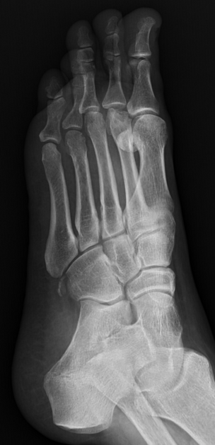
Freiberg disease, also known as Freiberg infraction, is osteochondrosis of the metatarsal heads. It commonly affects the 2nd or 3rdmetatarsal head (in ~2/3 and ~1/4 of all cases, respectively) or rarely, the 4th or 5th metatarsal head 13. It can be bilateral in up to 10% of cases.
On this page:
Epidemiology
Freiberg disease is most common in adolescent athletic females (M:F = 1:5) and young women. It is the fourth most common osteochondrosis and is the only osteochondrosis more common in females 13.
Clinical presentation
Patients typically present with localised forefoot plantar pain, especially on weight-bearing, with swelling and tenderness.
Pathology
The cause of Freiberg disease is controversial and is probably multifactorial. A traumatic insult in the form of either acute or repetitive injury and vascular compromise, perhaps due to an elongated 2nd metatarsal, are the most popular theories. As it is more commonly seen in women, particularly during adolescence, high-heeled shoes have been postulated as a possible causative factor.
Microscopic appearance
Histologically, Freiberg disease is characterised by the collapse of the subchondral bone, osteonecrosis, and cartilaginous fissures 1.
Classification
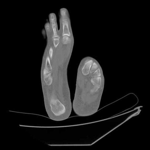
Some publications advocate the use of the Bragard staging classification 10, which requires two views/planes of the forefoot:
stage I: metatarsal head flattening and decreased subchondral bone density
stage II: metatarsal head sclerosis, fragmentation, and deformation, with cortical thickening
stage III: metatarsophalangeal osteoarthrosis with intra-articular loose bodies
Radiographic features
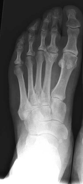
Plain radiograph
These can be split into early and late features:
Early
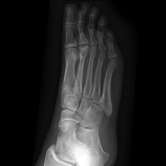
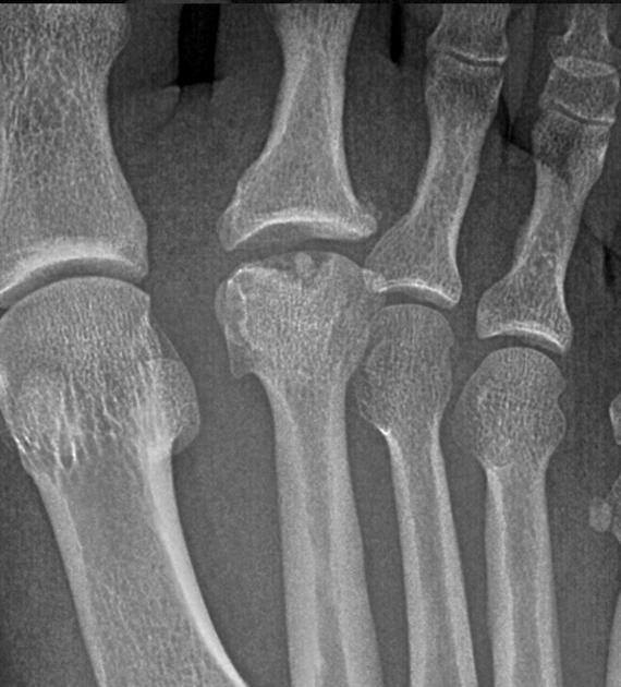
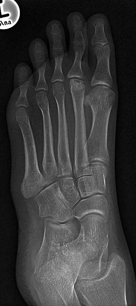
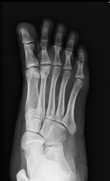
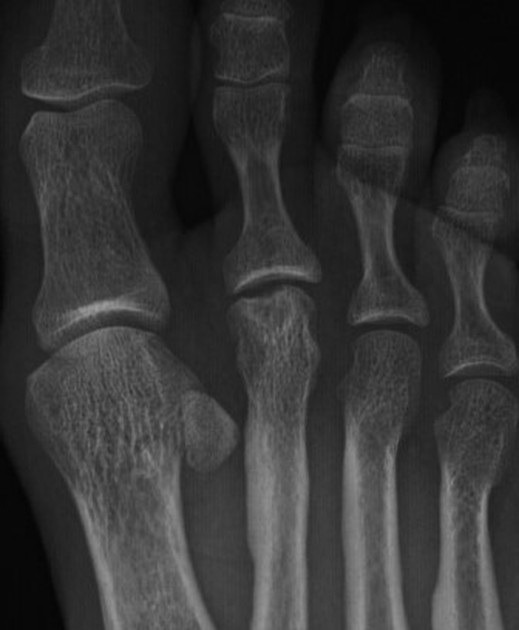
flattening and cystic lesions of the affected metatarsal head
widening of the metatarsophalangeal joint
Late
osteochondral fragments
sclerosis and flattening of the bone
increased cortical thickening
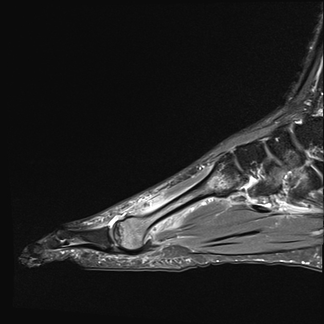
MRI
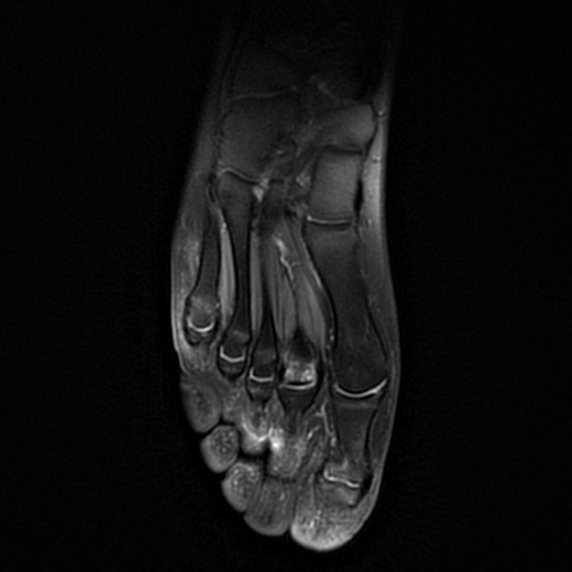
Early MRI findings include low-signal-intensity changes in the metatarsal head on T1-weighted images with increased signal intensity on corresponding T2-weighted and STIR images.
With disease progression, flattening of the metatarsal head occurs, and low-signal-intensity changes develop on T2-weighted images as the bone becomes sclerotic.
Treatment and prognosis
In early stages of the disease, it can be managed with lifestyle modifications, immobilisation and NSAID therapy. In advanced stages, surgery can be considered. It is usually divided into joint preserving or reconstruction options 12.
History and etymology
Albert H Freiberg (1868-1940) was an American orthopaedic surgeon who first described his eponymous condition in 1914 8,9,11.
Differential diagnosis
On imaging consider
normal variant: metatarsal head flattening is described in ~10% of the asymptomatic population
fracture of metatarsal head or neck including subchondral insufficiency fracture
lesser metatarsal head instability (only identified on MRI) due to plantar plate tear


 Unable to process the form. Check for errors and try again.