
Gallbladder ultrasound is a non-invasive diagnostic imaging technique used to evaluate the structure and function of the gallbladder as well as the adjacent anatomy.
On this page:
Preparation
Patients are typically advised to fast for 6-8 hours prior to the ultrasound examination. This allows the gallbladder to be adequately distended, thus enabling accurate assessment of the wall thickness and gallbladder contents. Fasting also allows for clearer visualisation of the gallbladder by reducing the volume of bowel gas and food contents within the gastrointestinal tract 1.
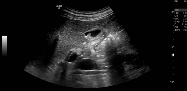
Techniques
supine position: the patient lies down on their back initially, allowing for general evaluation of the liver and gallbladder area; the ultrasound probe (transducer) should be placed just below the right ribcage, slightly angled towards the patient's head 2
intercostal scan: with the patient continuing in a supine position, an intercostal scan can be performed by placing the probe between ribs 8-11 in a sagittal plane; this helps assess potential lesions that might be obscured by rib shadowing in other views 3
subcostal scan: the patient remains supine while the probe is placed just under the right costal margin but more medial than before; this view provides better visualisation of neck and fundus regions of the gallbladder 4
lateral decubitus position: the patient lies on their left side, with the right arm raised above the head; the probe can be positioned along the right midaxillary line, with a slight oblique angle to optimise visualisation of the gallbladder 5
posterior oblique position: referred to as the "gallbladder easement" position, the patient lies down at a 45-degree angle, with their back supported and right arm overhead; this position allows visualisation of the gallbladder by bringing it closer to the abdominal wall and reducing gas interference 6
Indications
Gallbladder ultrasound is indicated in several clinical scenarios, including ref:
evaluation of right upper quadrant abdominal pain
suspected acute cholecystitis
suspected cholelithiasis
assessing complications related to cholelithiasis such as choledocholithiasis
evaluation of jaundice, particularly when a biliary cause is suspected
guidance for biopsy or drainage procedures
Normal findings
Normal findings on a gallbladder ultrasound include a thin-walled (<3 mm), anechoic, and pear-shaped structure that typically measures between 7–10 cm in length and 3–4 cm in width. The bile ducts should also appear anechoic without evidence of dilation or obstruction 7,8.
Related pathology
cholelithiasis: stones within the gallbladder, appearing as echogenic foci with posterior acoustic shadowing 9
acute cholecystitis: wall thickening (>3 mm), pericholecystic fluid, and sonographic Murphy’s sign (pain on probe compression) 10
chronic cholecystitis: wall thickening (>3mm) with fibrosis and occasional presence of gallstones 11
choledocholithiasis: stones in the extrahepatic bile ducts, can cause biliary obstruction 12
adenomyomatosis: Rokitansky-Aschoff sinuses with comet-tail artifacts, representing hypertrophy and invagination of the gallbladder wall 13
gallbladder polyps: small projections from the gallbladder wall without acoustic shadowing +/- vascularity 14
gallbladder carcinoma: rare but aggressive malignancy of the gallbladder characterised as irregular gallbladder wall thickening, invasion into adjacent structures, or metastatic lymph nodes in the surrounding area 15
Practical points
the sensitivity and specificity of ultrasound for detecting gallstones approach 96% and 90%, respectively 16
it is essential to perform a complete examination of the liver, bile ducts, and pancreas along with the gallbladder to detect any additional abnormalities


 Unable to process the form. Check for errors and try again.